Image-Guided Cancer Treatments in Vijayawada
Interventional oncology uses imaging to deliver treatment directly to selected tumours. Depending on the cancer, options can include transarterial chemoembolisation (TACE), radioembolisation, bland embolisation, thermal ablation, biopsy, drainage and venous access procedures.
Understanding Interventional Oncology
Cancer care is multidisciplinary. An interventional radiologist works with medical, surgical and radiation oncologists, hepatologists and other specialists. The treatment plan depends on the tumour type, stage, location, number and size of lesions, liver or kidney function and treatments already received.
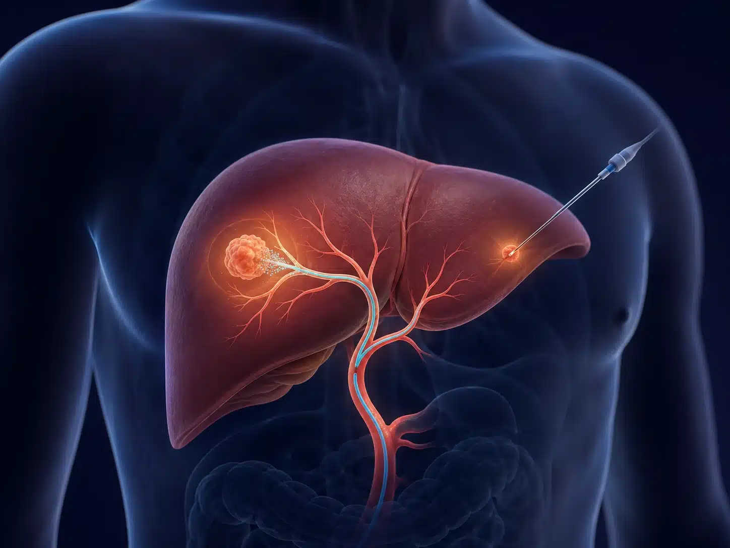
For liver tumours, embolisation delivers therapy through the hepatic artery and reduces blood flow to the target. Ablation places a needle into a selected tumour under ultrasound or CT guidance and uses heat or another energy to destroy tissue. These treatments may aim for local control, symptom relief, bridging to transplant or combination care.
Interventional oncology in Vijayawada should be offered only after review of high-quality imaging and pathology when required. It is not a replacement for every surgery, chemotherapy or radiation plan. Clear goals, expected benefits, limitations and follow-up imaging are discussed before treatment.
Understanding the condition and treatment pathway
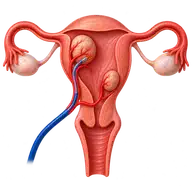
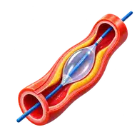
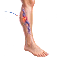
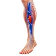
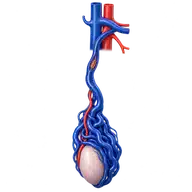
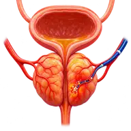
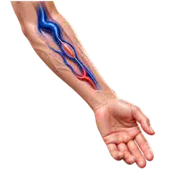
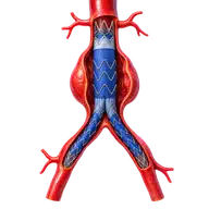
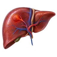
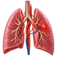
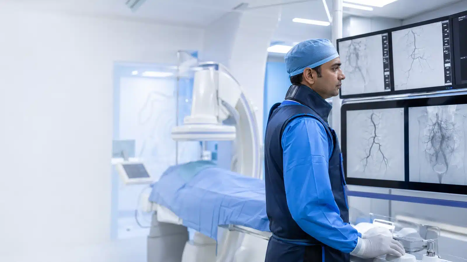
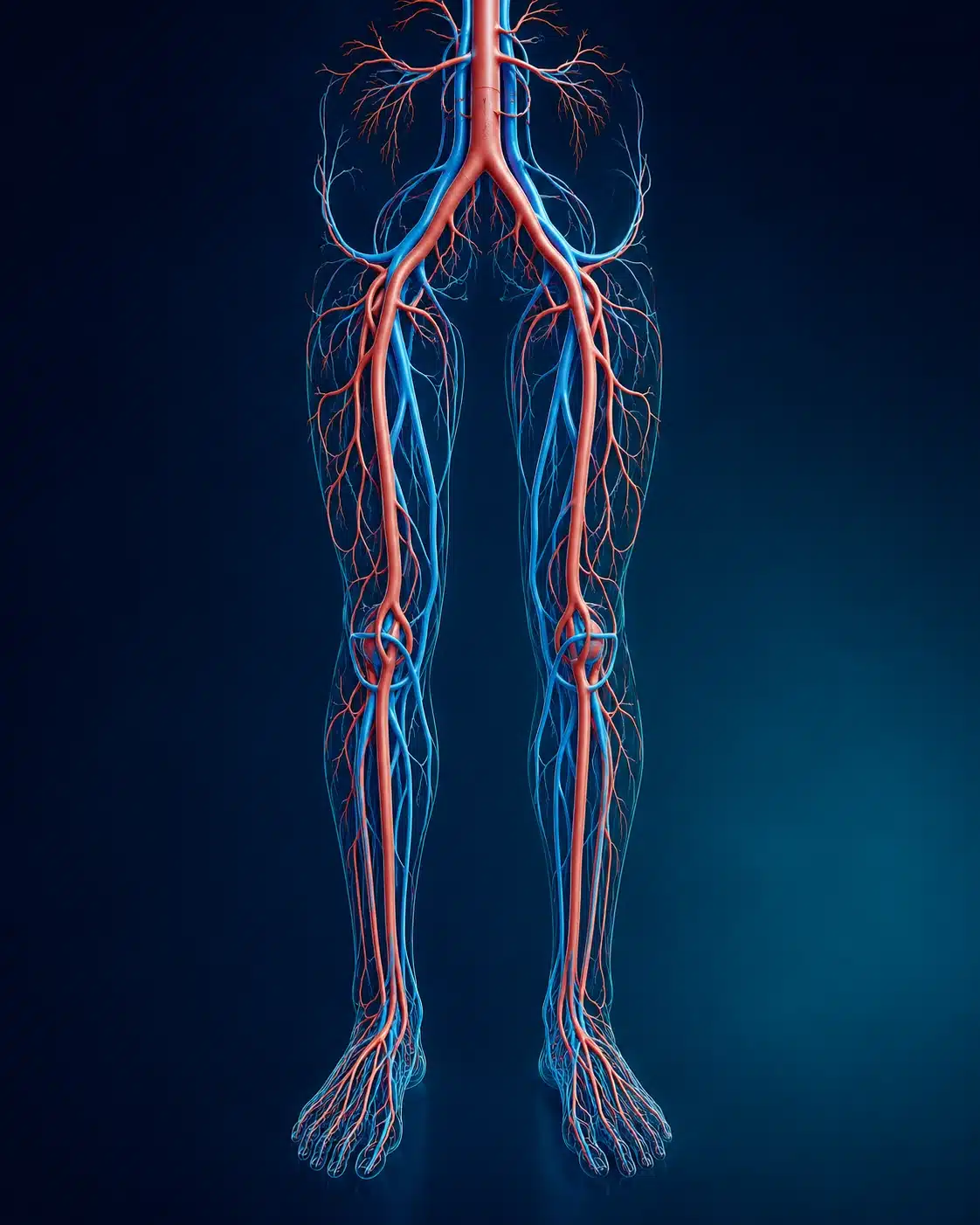
These medical visuals show the anatomy, treatment target and image-guided route involved in Interventional Oncology . The actual procedure plan is based on the patient’s scans, symptoms and overall health.
Signs and symptoms
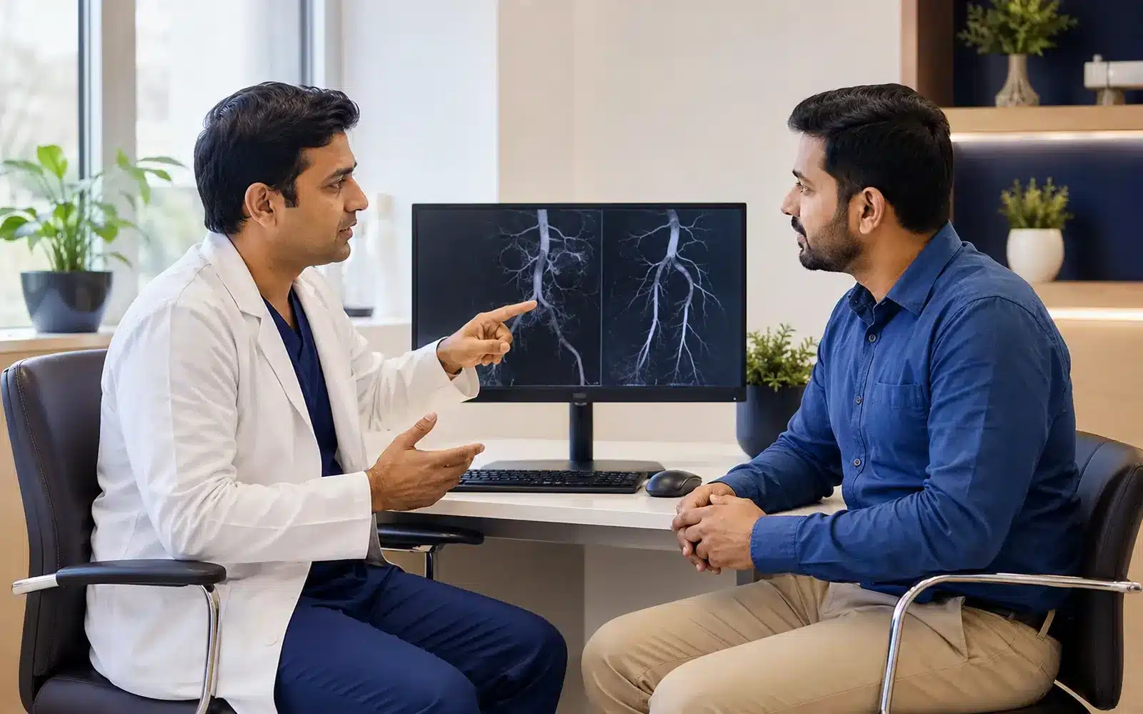
Evaluation & planning
A precise, image-guided pathway
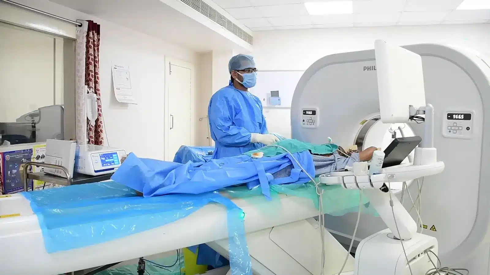
During Interventional Oncology , imaging helps the interventional radiologist follow the anatomy, position the catheter or needle and confirm the treatment target. The exact technique and devices are chosen for the patient’s diagnosis and scan findings.
The tumour and treatment route are planned from imaging.
Catheter or needle access is placed under image guidance.
Embolisation or ablation is delivered to the target.
Post-treatment imaging and follow-up assess response.
Recovery and follow-up
Fatigue, fever, pain or nausea may occur depending on treatment. Liver and kidney function, blood counts and access sites are monitored. The oncology team plans follow-up scans and any additional therapy.
Before discharge, the team explains medicines, activity, wound or access-site care, expected symptoms and the warning signs that should prompt urgent review. Follow-up timing depends on the condition and may include Doppler, CT, MRI, blood tests or consultation with another specialist.
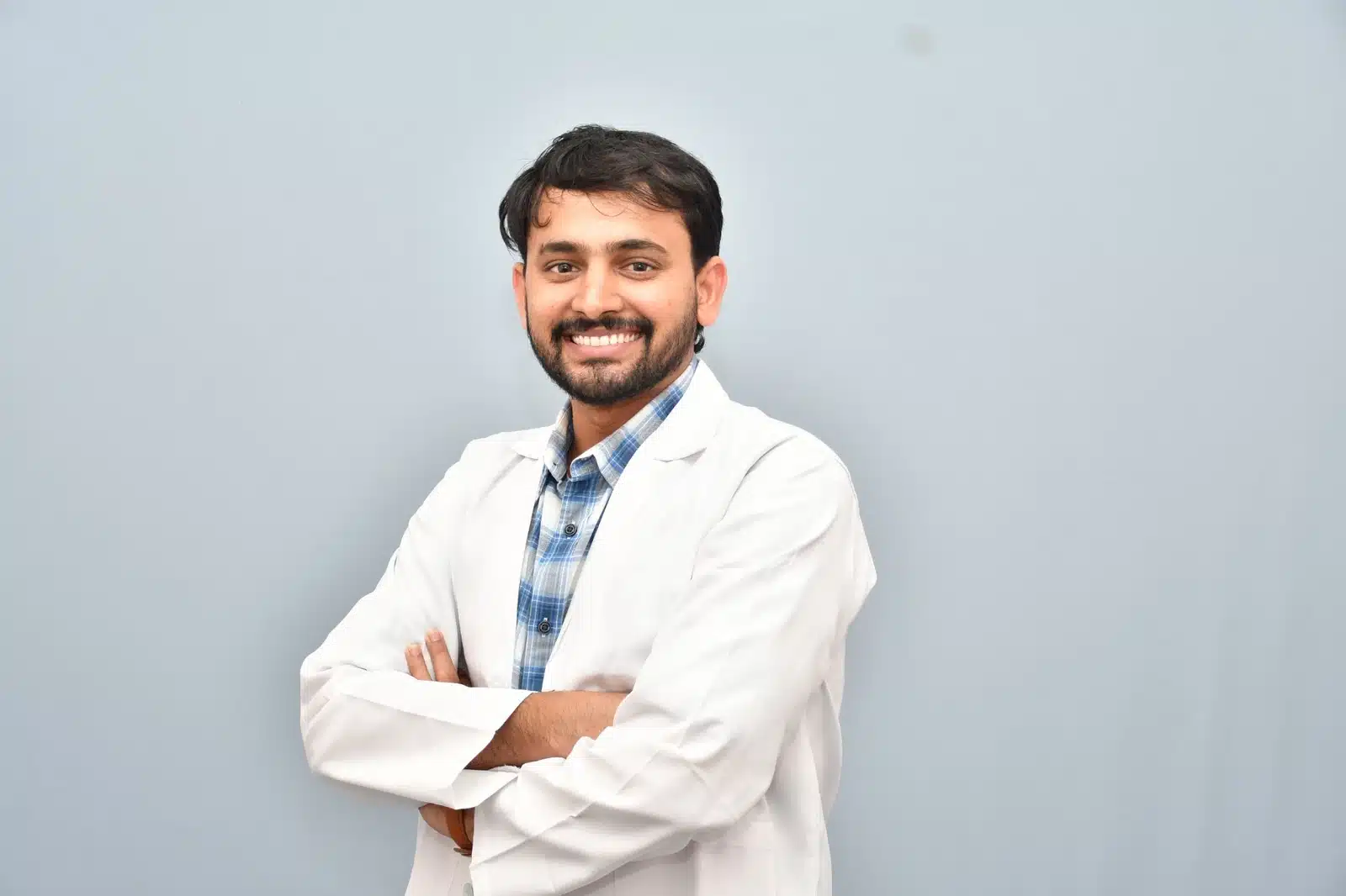
Meet Dr. M. Bhavani Shankar Manam
MBBS, MD, DNB (Radio Diagnosis), FIVR
Consultant Vascular & Interventional Radiologist with experience across vascular, non-vascular and oncological image-guided procedures and more than 3,000 cases. Every treatment plan begins with a review of symptoms, reports and imaging, followed by a clear discussion of suitable options.
Frequently asked questions
These answers are general and cannot determine your suitability without clinical review.
TACE combines local chemotherapy with arterial embolisation; other embolisation methods may not use chemotherapy.
For selected small tumours it may be an alternative, but surgery or systemic therapy may be better for others.
Follow-up contrast CT or MRI and clinical review assess treated tissue and remaining disease.