Varicocele Embolization in Vijayawada
Varicocele treatment in Vijayawada may be considered for persistent scrotal discomfort or selected fertility concerns after examination and appropriate tests. Varicocele embolization treats abnormal backward flow in the veins around the testicle. A vascular and interventional radiologist guides a fine catheter through a vein and closes the refluxing pathway using suitable embolic materials, without a scrotal incision.
Understanding Varicocele Embolization
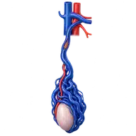
A varicocele is an enlargement of veins around the testicle, more often on the left side. It may cause a dragging ache, heaviness, visible enlarged veins, testicular size difference or an abnormal semen analysis. Many varicoceles need no intervention; treatment is considered after urology review when symptoms or fertility concerns are meaningful.
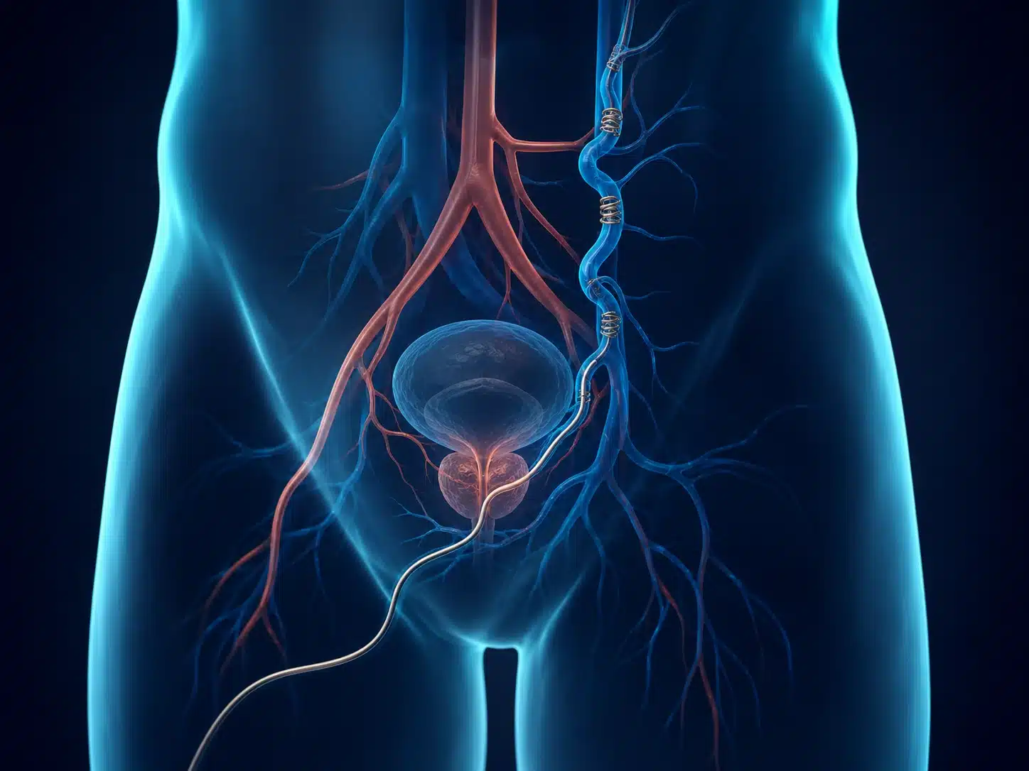
Varicocele embolization in Vijayawada avoids a scrotal incision. Venography maps the refluxing veins, including accessory channels that may contribute to recurrence. The abnormal pathway is then closed so blood redirects through healthy veins.
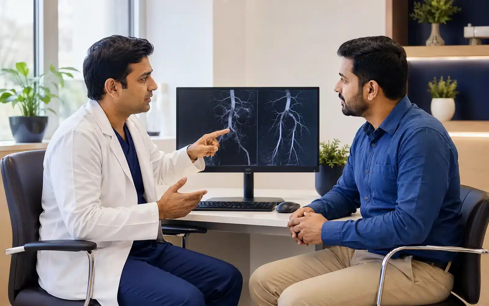
A varicocele grade alone does not decide whether treatment is needed. Symptoms, examination, Doppler findings, testicular size and semen analysis may influence the recommendation. Observation, surgical repair and embolization should be compared with the urologist. An abnormal semen analysis can have several causes, and varicocele treatment does not guarantee pregnancy. Bring previous ultrasound reports, semen tests and details of any earlier repair to the consultation.
Understanding the condition and treatment pathway
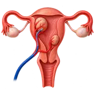
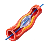
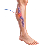
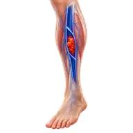
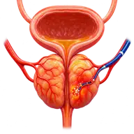
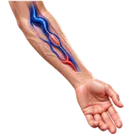
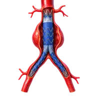
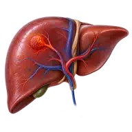
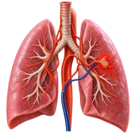
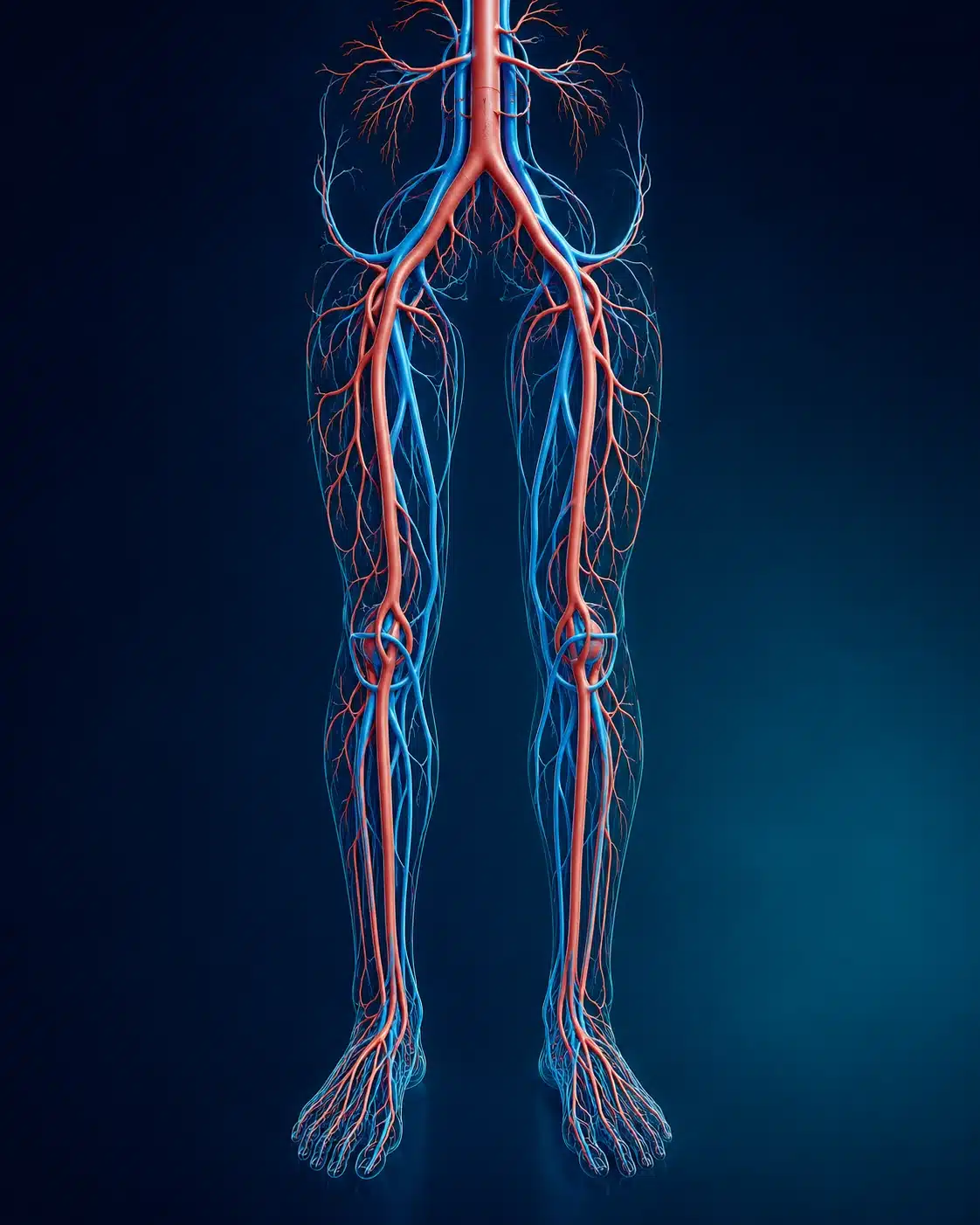
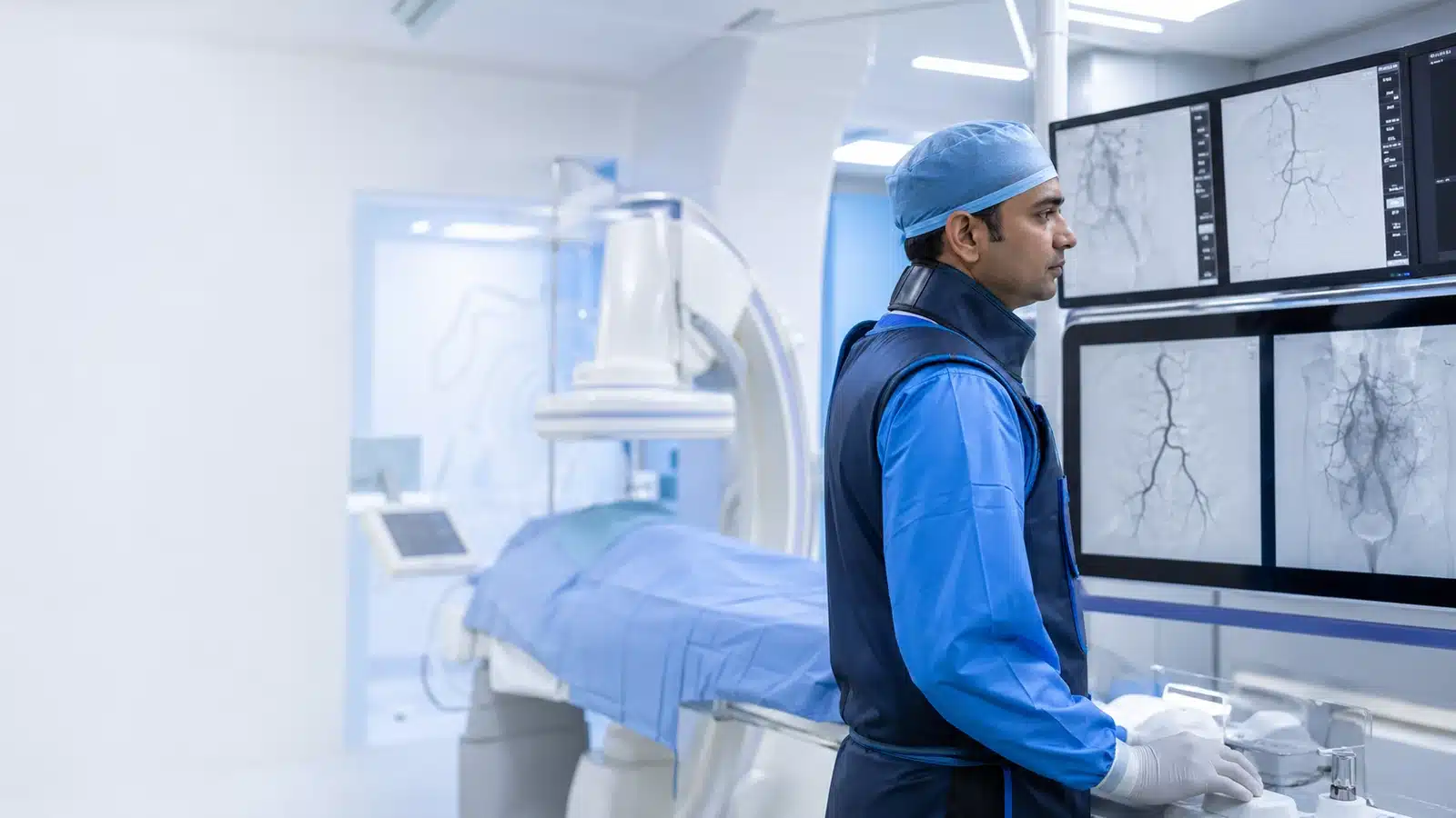
These medical visuals show the anatomy, treatment target and image-guided route involved in Varicocele Embolization . The actual procedure plan is based on the patient’s scans, symptoms and overall health.
Signs and symptoms
Evaluation & planning
A precise, image-guided pathway
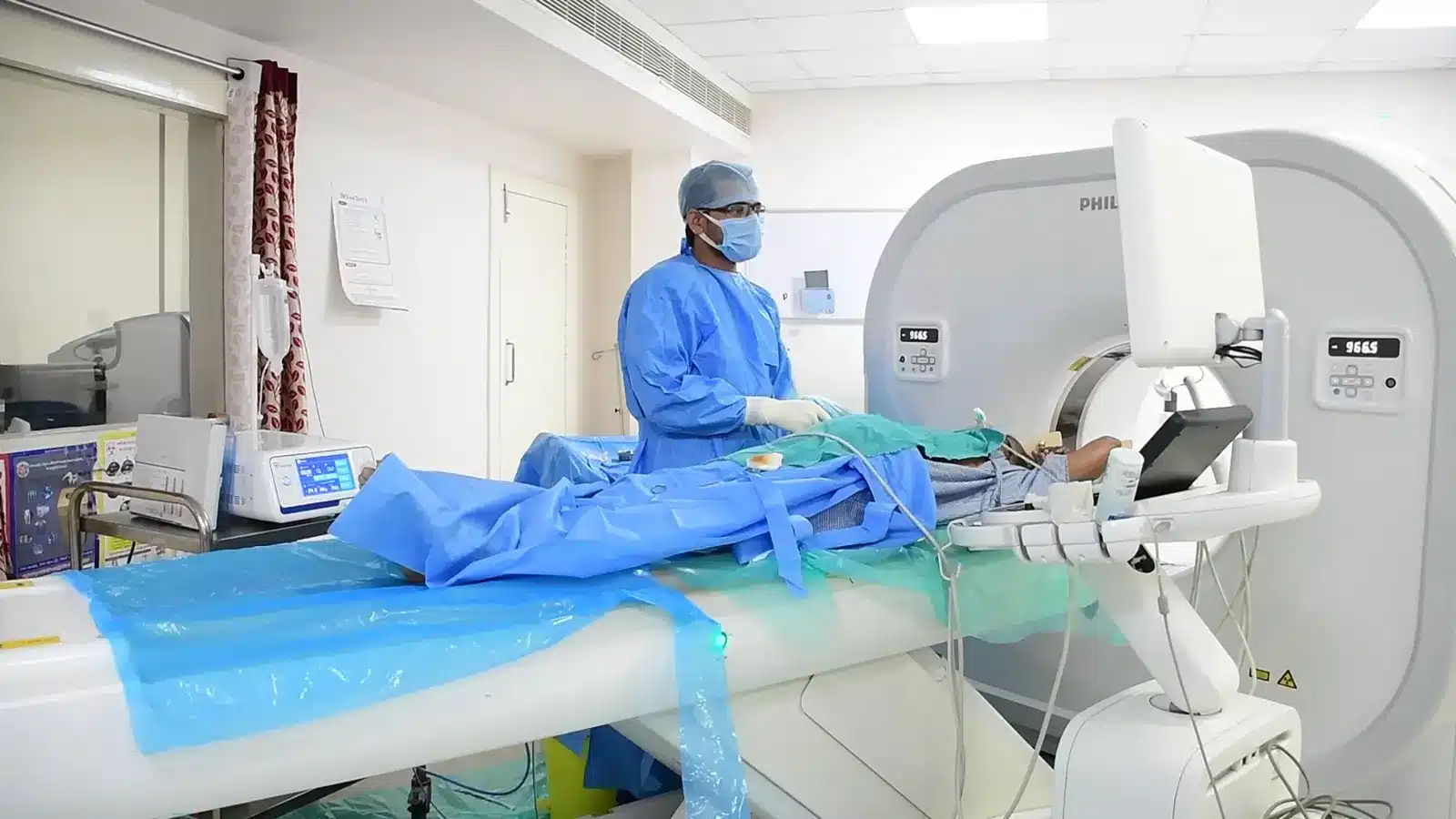
During Varicocele Embolization , imaging helps the interventional radiologist follow the anatomy, position the catheter or needle and confirm the treatment target. The exact technique and devices are chosen for the patient’s diagnosis and scan findings.
A small venous access point is prepared.
A fine catheter is guided to the gonadal vein.
Venography identifies reflux and side branches.
Coils, plugs or sclerosant close the abnormal vein.
Recovery and follow-up
Mild back, groin or scrotal discomfort can occur temporarily. Patients receive guidance about exercise, lifting, medicines and follow-up. Fertility assessment, if required, takes place over subsequent months.
After varicocele embolization, the team explains puncture-site care and when to resume lifting, exercise and sexual activity. Risks include bruising, infection, contrast reaction, persistent or recurrent varicocele and, rarely, migration of embolic material. Sometimes vein anatomy prevents catheter treatment. Follow-up checks symptoms and, when fertility is relevant, repeat semen testing over subsequent months. Sudden severe testicular pain needs urgent assessment rather than waiting for a routine appointment. Meet the vascular and interventional radiologist or arrange a report review.
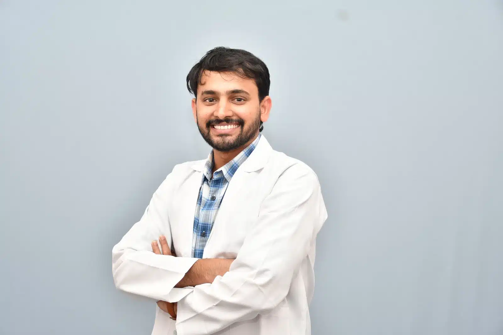
Meet Dr. M. Bhavani Shankar Manam
MBBS, MD, DNB (Radio Diagnosis), FIVR
Consultant Vascular & Interventional Radiologist with experience across vascular, non-vascular and oncological image-guided procedures and more than 3,000 cases. Every treatment plan begins with a review of symptoms, reports and imaging, followed by a clear discussion of suitable options.
Frequently asked questions
These answers are general and cannot determine your suitability without clinical review.
It may improve semen parameters in selected men, but pregnancy cannot be guaranteed and both partners may need evaluation.
Small medical coils or plugs may remain in the treated vein to keep it closed.
Recurrence is possible after any treatment. Imaging helps identify accessory veins and plan management.