Varicose Veins Treatment in Vijayawada
Varicose veins treatment in Vijayawada begins with a clinical examination and venous Doppler scan to identify the source of reflux. Depending on the vein pattern and symptoms, care may include compression, endovenous laser or radiofrequency ablation, sclerotherapy or small-vein removal.
Understanding Varicose Veins
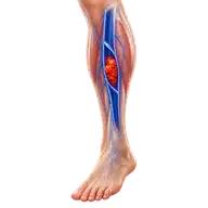
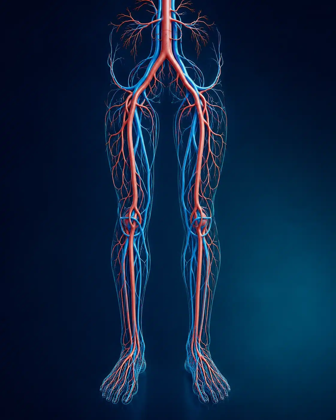
Varicose veins are enlarged, twisted surface veins caused by weak or poorly functioning valves. Symptoms may include aching, heaviness, ankle swelling, itching, night cramps and burning. Advanced venous disease can cause skin darkening, eczema, hardening around the ankle, bleeding or a venous ulcer.
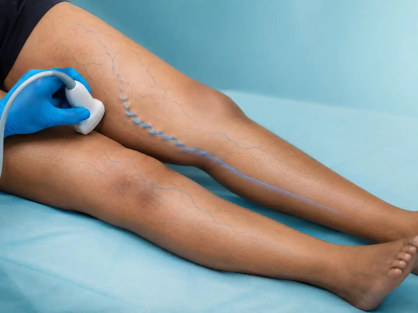
A visible vein is only part of the story. Doppler ultrasound maps deep and superficial veins, checks valve function and looks for clot. This helps avoid one-size-fits-all treatment. Small spider veins, a leaking main superficial vein and recurrent veins after surgery require different plans.
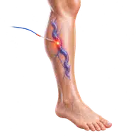
Endovenous ablation closes an unhealthy superficial vein from inside using controlled heat. Blood naturally returns through healthier veins. Sclerotherapy can treat selected branch or spider veins, while ambulatory phlebectomy removes prominent surface veins through tiny punctures. Compression and movement remain important before and after treatment when advised.
Understanding the condition and treatment pathway
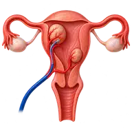
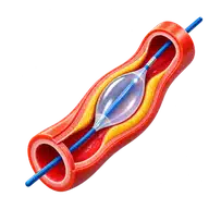
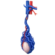
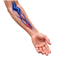
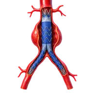
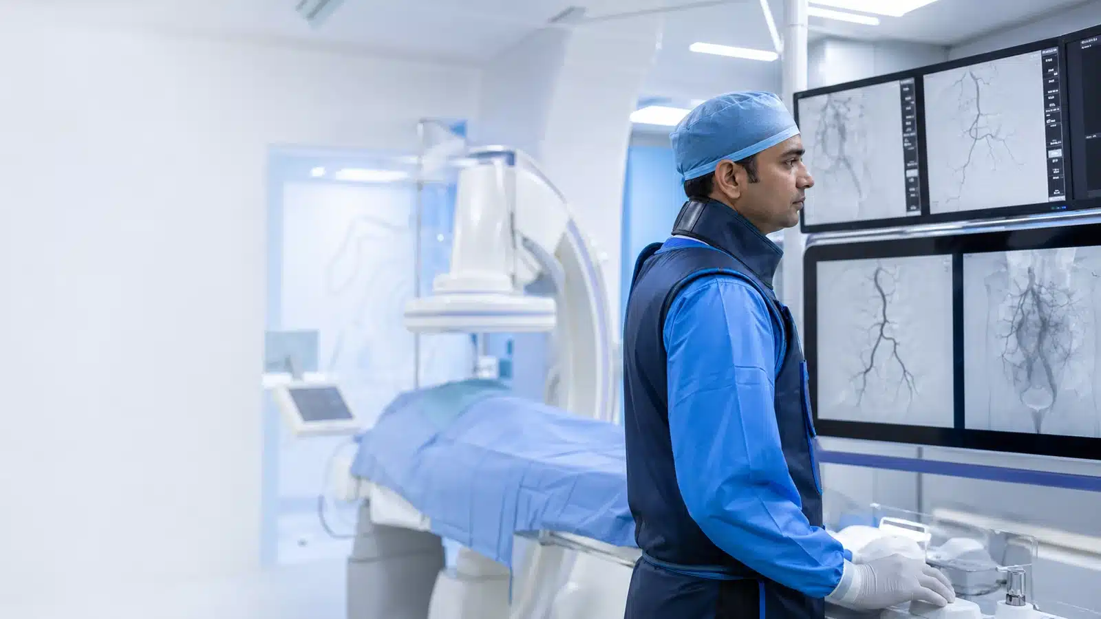
These medical visuals show the anatomy, treatment target and image-guided route involved in Varicose Veins . The actual procedure plan is based on the patient’s scans, symptoms and overall health.
Signs and symptoms
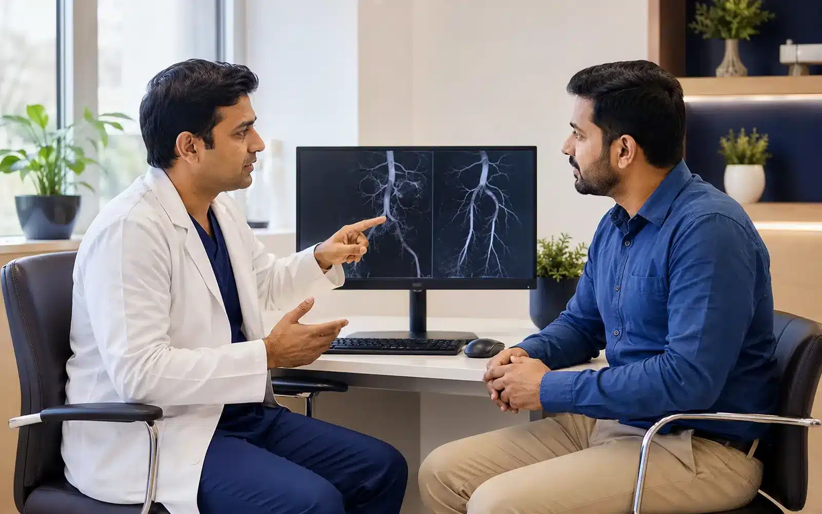
Evaluation & planning
A precise, image-guided pathway
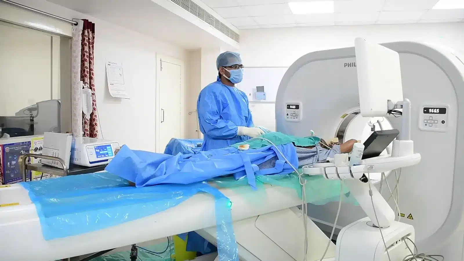
During Varicose Veins , imaging helps the interventional radiologist follow the anatomy, position the catheter or needle and confirm the treatment target. The exact technique and devices are chosen for the patient’s diagnosis and scan findings.
The target vein is mapped with ultrasound.
Local anaesthetic is used around the treatment area.
Laser, radiofrequency, glue, foam or another selected method closes the unhealthy vein.
Walking and aftercare begin as advised.
Recovery and follow-up
Temporary tightness, bruising or tenderness can occur. The doctor explains walking, compression, bathing, travel and medicine instructions. Follow-up Doppler may be advised based on the procedure and symptoms.
Varicose vein treatment planning considers symptoms, Doppler findings, skin changes and previous procedures. Ask about the suitable technique, compression advice, follow-up visits and the total cost of care rather than choosing by a laser-treatment label alone. Risks include bruising, inflammation, skin or nerve injury and uncommon blood clots; veins can recur. Sudden one-sided swelling requires assessment for DVT. Bleeding from a vein or a worsening leg ulcer needs prompt care. Bring earlier Doppler scans to the vascular consultation.
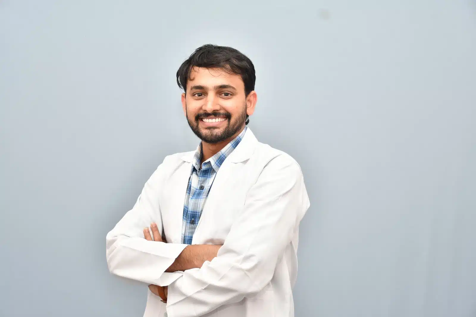
Meet Dr. M. Bhavani Shankar Manam
MBBS, MD, DNB (Radio Diagnosis), FIVR
Consultant Vascular & Interventional Radiologist with experience across vascular, non-vascular and oncological image-guided procedures and more than 3,000 cases. Every treatment plan begins with a review of symptoms, reports and imaging, followed by a clear discussion of suitable options.
Frequently asked questions
These answers are general and cannot determine your suitability without clinical review.
Unhealthy superficial veins can be closed while blood returns through healthy deep and superficial pathways confirmed on Doppler.
Not always. Some veins reduce over time and some need additional sclerotherapy or phlebectomy.
New reflux or branch veins can develop later, so follow-up and risk reduction remain useful.