Fistuloplasty for Dialysis Access in Vijayawada
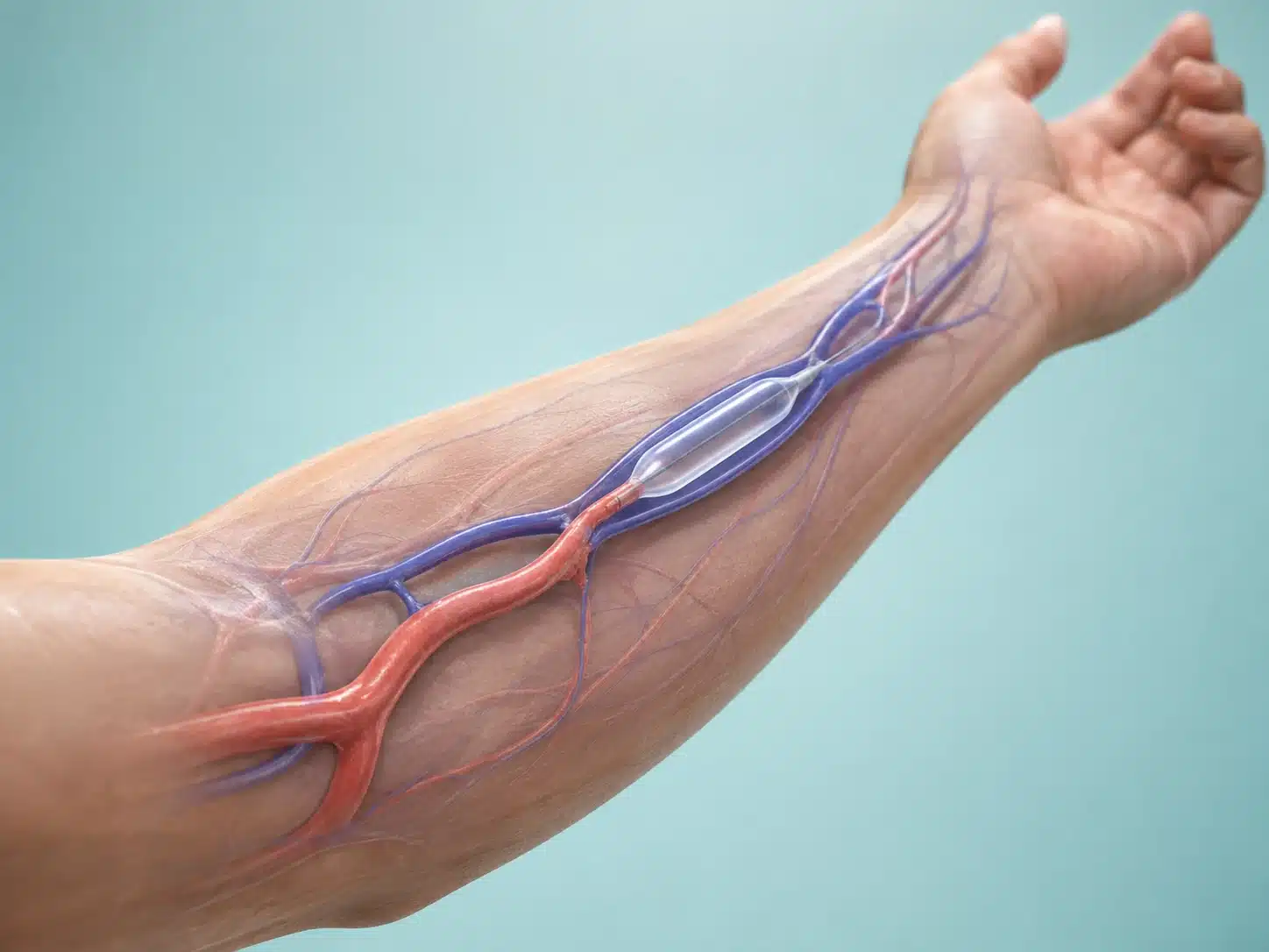
Fistuloplasty is an angioplasty performed to widen a narrowed dialysis fistula or graft. By improving blood flow through the access, it may support effective dialysis and help preserve an access that is becoming difficult to use.
Understanding Fistuloplasty
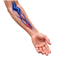
An arteriovenous fistula is created surgically for haemodialysis. Over time, narrowing can develop in the inflow artery, the fistula or the draining vein. Warning signs include low dialysis flow, high venous pressure, difficult needle placement, prolonged bleeding, arm swelling or a change in the normal buzzing sensation.
Fistuloplasty in Vijayawada uses ultrasound and X-ray guidance. Contrast imaging identifies the narrowed segment. A balloon is inflated across the stenosis, and a stent may be considered in specific recurrent or resistant lesions. Fresh clot may require additional thrombectomy or thrombolysis.
Early referral can improve the chance of preserving the access. A completely failed or infected access may require a different plan involving nephrology, vascular surgery and the dialysis team.
Understanding the condition and treatment pathway
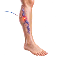
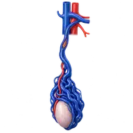
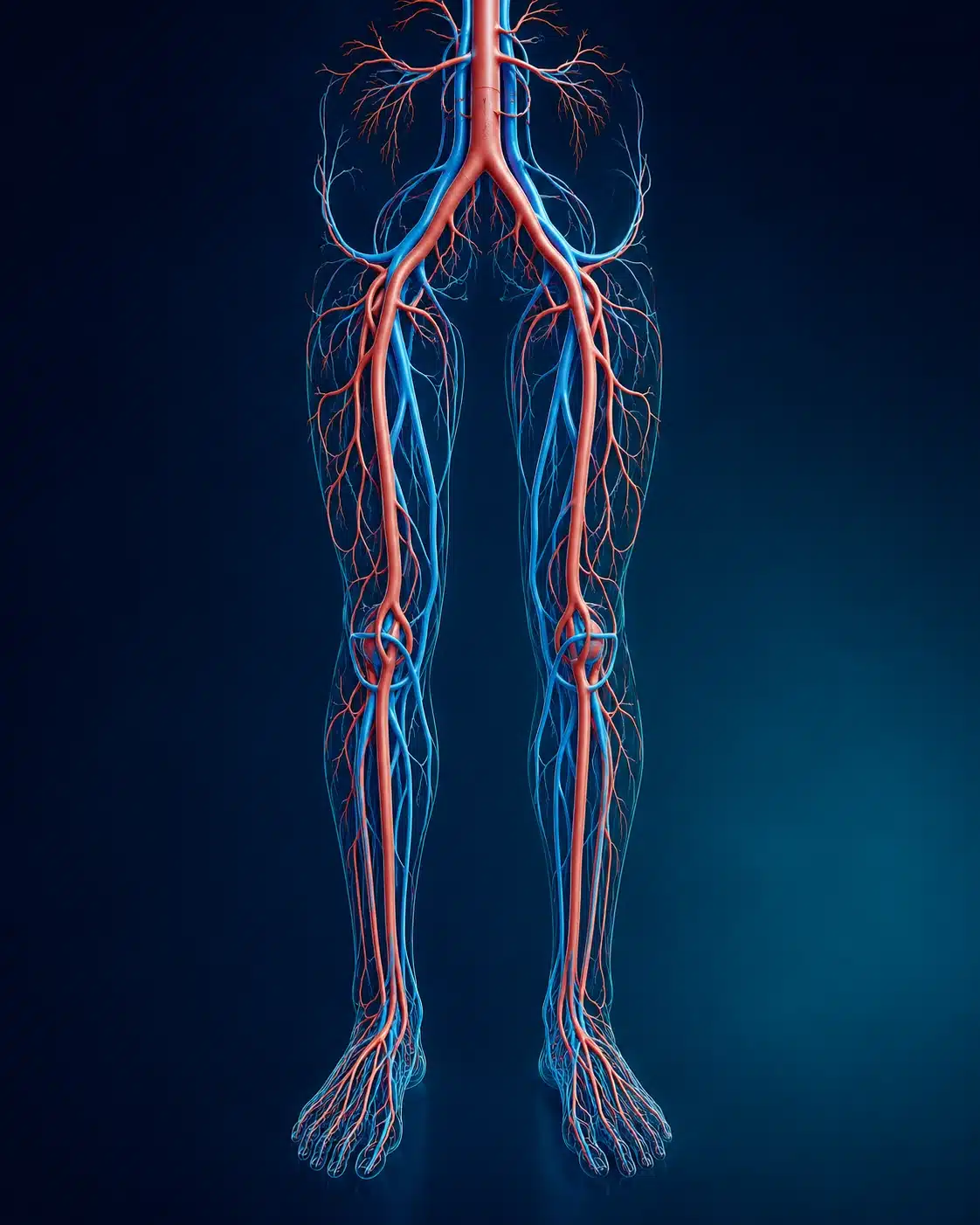
These medical visuals show the anatomy, treatment target and image-guided route involved in Fistuloplasty . The actual procedure plan is based on the patient’s scans, symptoms and overall health.
Signs and symptoms
Evaluation & planning
A precise, image-guided pathway
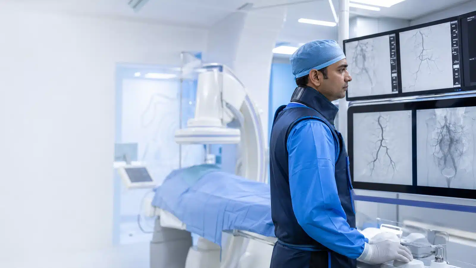
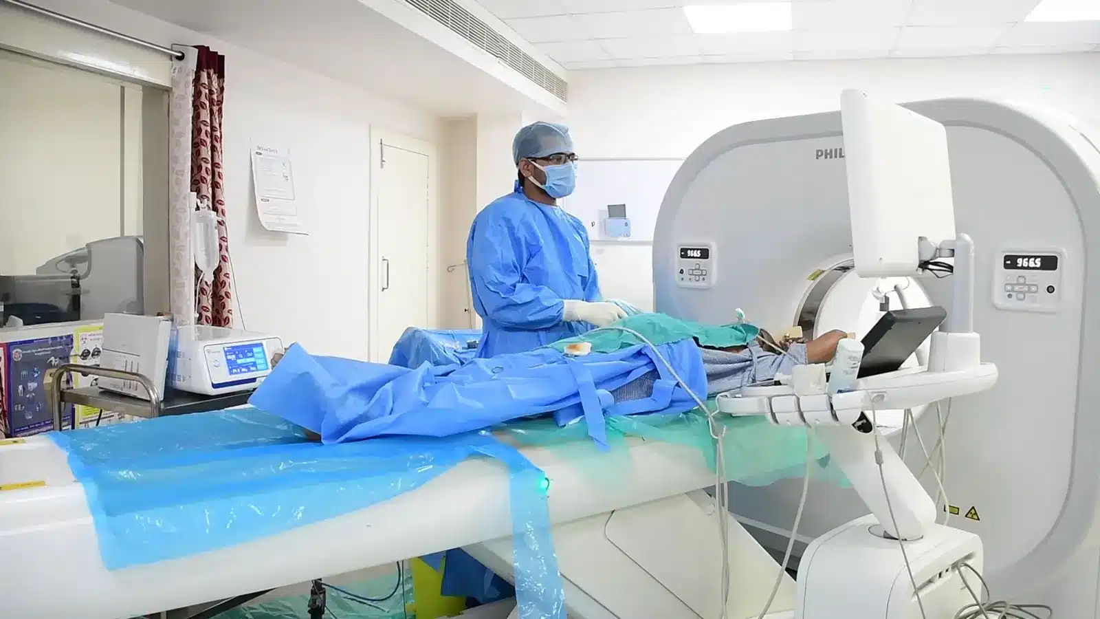
During Fistuloplasty , imaging helps the interventional radiologist follow the anatomy, position the catheter or needle and confirm the treatment target. The exact technique and devices are chosen for the patient’s diagnosis and scan findings.
The access is entered through a small puncture.
A fistulogram maps the narrowing.
A balloon widens the affected segment.
Flow is rechecked and further treatment is used if needed.

Recovery and follow-up
The thrill and circulation are checked after treatment. Patients receive access-care, bleeding and dialysis instructions. Re-narrowing can occur, so dialysis-unit surveillance remains important.
Before discharge, the team explains medicines, activity, wound or access-site care, expected symptoms and the warning signs that should prompt urgent review. Follow-up timing depends on the condition and may include Doppler, CT, MRI, blood tests or consultation with another specialist.

Meet Dr. M. Bhavani Shankar Manam
MBBS, MD, DNB (Radio Diagnosis), FIVR
Consultant Vascular & Interventional Radiologist with experience across vascular, non-vascular and oncological image-guided procedures and more than 3,000 cases. Every treatment plan begins with a review of symptoms, reports and imaging, followed by a clear discussion of suitable options.
Frequently asked questions
These answers are general and cannot determine your suitability without clinical review.
Many accesses can be used soon after treatment, but timing is decided for the individual access.
Yes. Repeat treatment may be required because dialysis access vessels can re-narrow.
No. Balloon angioplasty is usual; stents are reserved for selected situations.
You may also want to understand
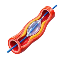
Peripheral Angioplasty
Peripheral angioplasty is a minimally invasive treatment used in selected patients to reopen narrowed or blocked arteries, mo …
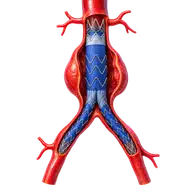
Aorta Stenting
Aortic stent-graft repair is a minimally invasive treatment for selected aortic aneurysms, dissections and injuries. A fabric …
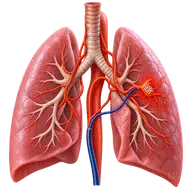
Bronchial Artery Embolisation
Bronchial artery embolisation (BAE) is an image-guided emergency or planned treatment used to control significant or recurren …