Pulmonary artery pseudoaneurysm with coughing blood
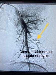
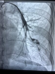
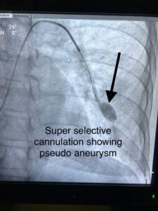
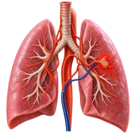
The clinic’s case report describes a 73-year-old woman with tuberculosis who developed episodes of massive haemoptysis, or coughing up blood. Imaging identified a large pseudoaneurysm in a segmental pulmonary artery in the left lower lobe. This was assessed as the source of bleeding.
Assessment and embolisation
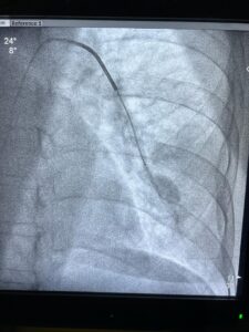
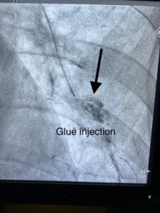
A pseudoaneurysm is a contained leak through a damaged artery wall. In this case, imaging was used to locate the abnormal vessel and plan embolisation. A catheter was guided through the blood vessels under imaging, and embolic material was used to block flow into the pseudoaneurysm.
Pulmonary artery embolisation and bronchial artery embolisation target different blood supplies. Identifying the actual bleeding source is important when planning treatment for haemoptysis.
Reported outcome and follow-up
The original report records no further haemoptysis after treatment, improvement during observation and discharge with follow-up instructions. These are the reported findings for this patient, rather than a prediction of another person’s outcome.
What patients should know
Heavy coughing of blood, breathlessness or faintness needs emergency hospital care. Embolisation carries risks including vessel injury, bleeding, contrast reactions and unintended blockage of other vessels. Treatment of the underlying lung condition and follow-up remain important.
For planned vascular review in Vijayawada, bring CT images and previous lung treatment records. Read the haemoptysis and BAE treatment guide for related information, or contact the clinic for a non-emergency appointment.