Embolization for Gastrointestinal Bleeding in Vijayawada
Transcatheter embolization can control selected gastrointestinal bleeding when endoscopy is unsuccessful, not possible or when CT shows an arterial source. The procedure targets the bleeding artery through a catheter while trying to preserve circulation to healthy bowel.
Gastrointestinal bleeding can present as vomiting blood, black stools, maroon stools, fresh rectal bleeding, dizziness, weakness or low blood pressure. Causes include ulcers, tumours, diverticular bleeding, post-surgical bleeding, vascular lesions and pseudoaneurysms.
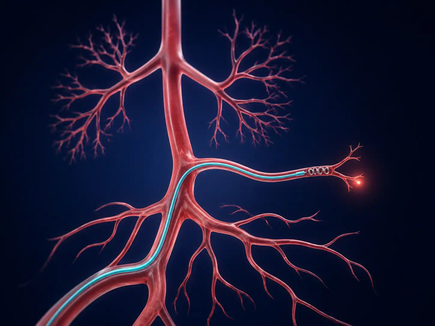
In GI bleeding embolization in Vijayawada, angiography searches for contrast leak or an abnormal artery. A microcatheter is advanced close to the source and coils, particles, liquid embolic or another device is selected for the anatomy. Endoscopy, surgery and interventional radiology work together.
The procedure is often performed urgently after stabilisation with fluids, blood products and medicines. The team balances bleeding control against the risk of reducing blood flow to bowel or other organs.
Understanding the condition and treatment pathway
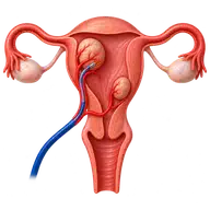
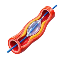
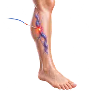
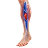
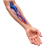
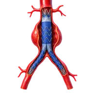
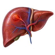
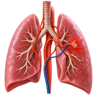
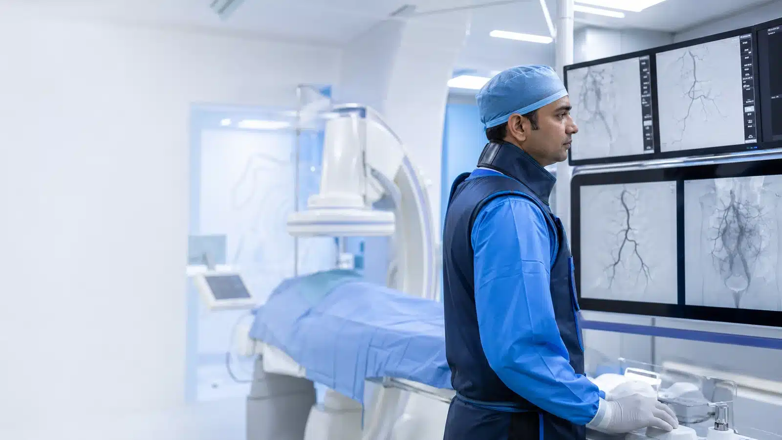
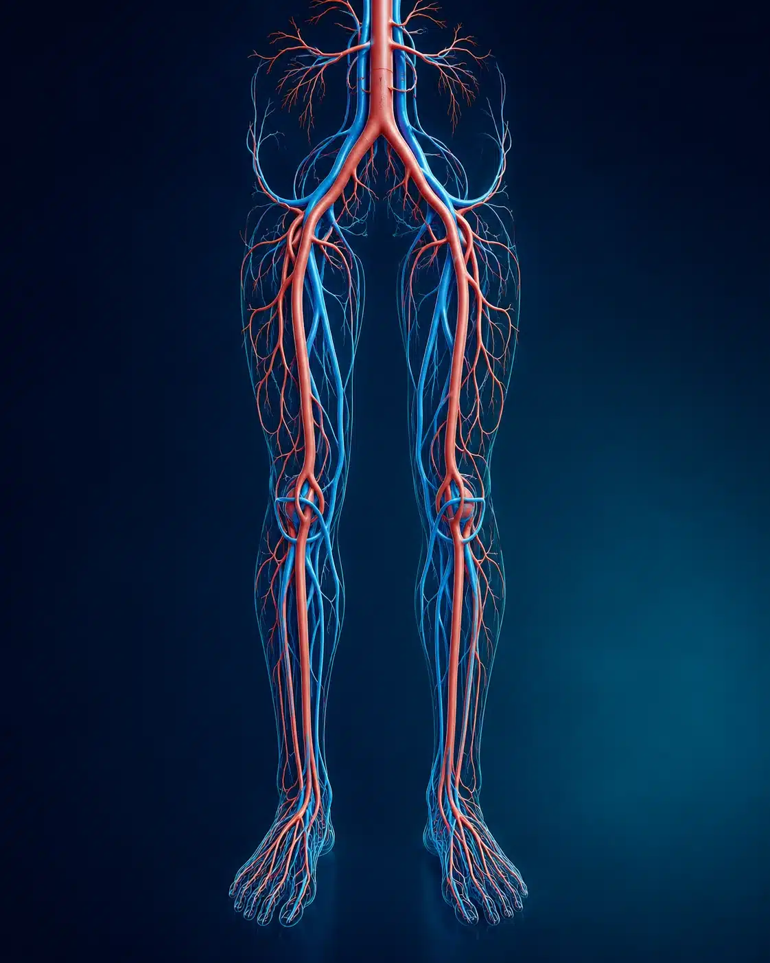
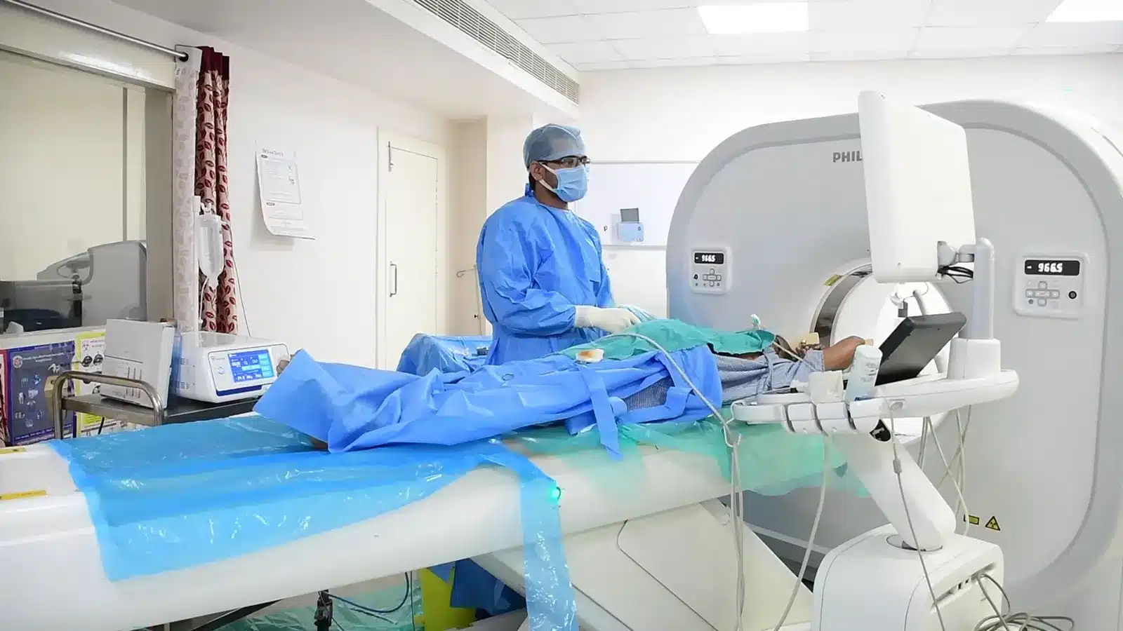
These medical visuals show the anatomy, treatment target and image-guided route involved in GI Bleeding Embolization. The actual procedure plan is based on the patient’s scans, symptoms and overall health.
Signs and symptoms
✓
Vomiting blood or coffee-ground material
✓
Black, maroon or bloody stool
✓
Faintness, fast pulse or low blood pressure
✓
Falling haemoglobin or transfusion need
Evaluation & planning
✓
Emergency stabilisation and blood tests
✓
Endoscopy when appropriate
✓
CT angiography
✓
Review of blood thinners and clotting
A precise, image-guided pathway
During GI Bleeding Embolization, imaging helps the interventional radiologist follow the anatomy, position the catheter or needle and confirm the treatment target. The exact technique and devices are chosen for the patient’s diagnosis and scan findings.
01
Arterial access is obtained.
02
Angiography identifies or localises the source.
03
A microcatheter reaches the target branch.
04
Embolic material seals the bleeding vessel.
Recovery and follow-up
Vitals, haemoglobin, abdominal symptoms and access site are monitored. Rebleeding may require repeat endoscopy, embolisation or surgery. New severe abdominal pain needs urgent review.
Before discharge, the team explains medicines, activity, wound or access-site care, expected symptoms and the warning signs that should prompt urgent review. Follow-up timing depends on the condition and may include Doppler, CT, MRI, blood tests or consultation with another specialist.
Consultant Vascular & Interventional Radiologist with experience across vascular, non-vascular and oncological image-guided procedures and more than 3,000 cases. Every treatment plan begins with a review of symptoms, reports and imaging, followed by a clear discussion of suitable options.