Bronchial Artery Embolisation for Coughing Blood in Vijayawada
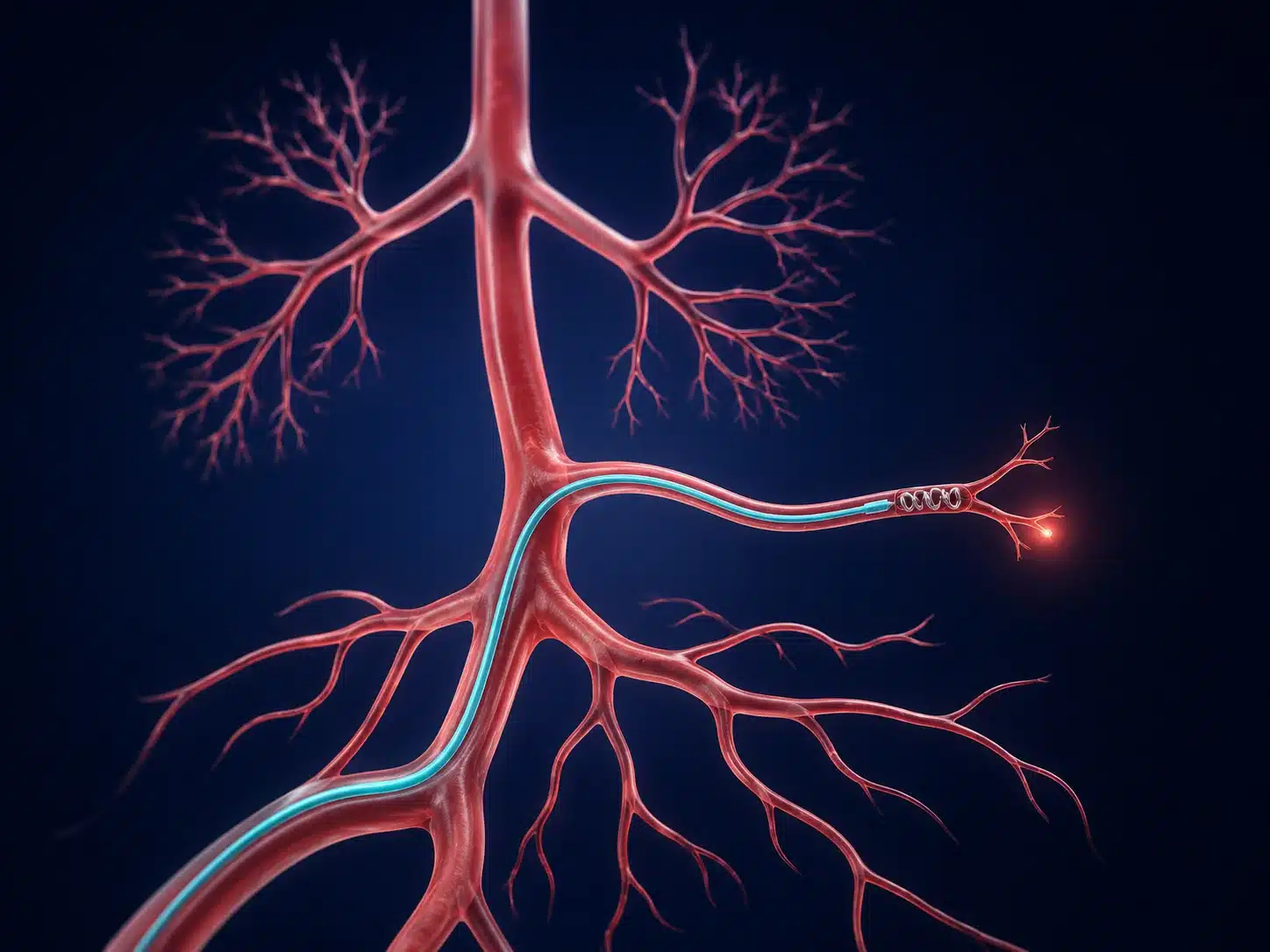
Bronchial artery embolisation in Vijayawada is an image-guided option for selected patients with significant or recurrent coughing of blood, called haemoptysis. A vascular and interventional radiologist uses a fine catheter to reach abnormal arteries supplying the affected lung area and block the bleeding source. Breathing difficulty, ongoing heavy bleeding or faintness requires emergency assessment rather than waiting for an appointment.
Understanding Bronchial Artery Embolisation
Coughing blood, called haemoptysis, can occur with infection, bronchiectasis, tuberculosis-related lung damage, tumours, vascular abnormalities and other conditions. Even a smaller amount can become urgent when bleeding continues, breathing is affected or the patient is medically fragile.
Bronchial artery embolisation in Vijayawada is planned using CT and angiography. The interventional radiologist maps arteries supplying the abnormal lung area while carefully identifying branches that must be protected. The goal is to control bleeding; the underlying lung disease still requires treatment.
Assessment may involve chest CT, blood tests and bronchoscopy, depending on the patient’s stability and the suspected cause. Bring previous tuberculosis treatment records, chest scans and a medicine list when available, but do not delay urgent care to collect reports. BAE controls bleeding from selected arteries; it does not remove the underlying lung disease. Respiratory specialists continue treatment for infection, bronchiectasis, tumours or other causes. Rebleeding may require another procedure or a different treatment.
Understanding the condition and treatment pathway
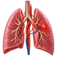
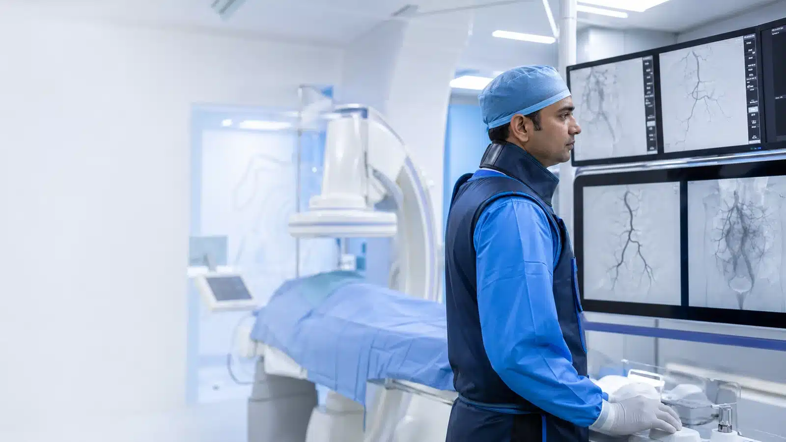
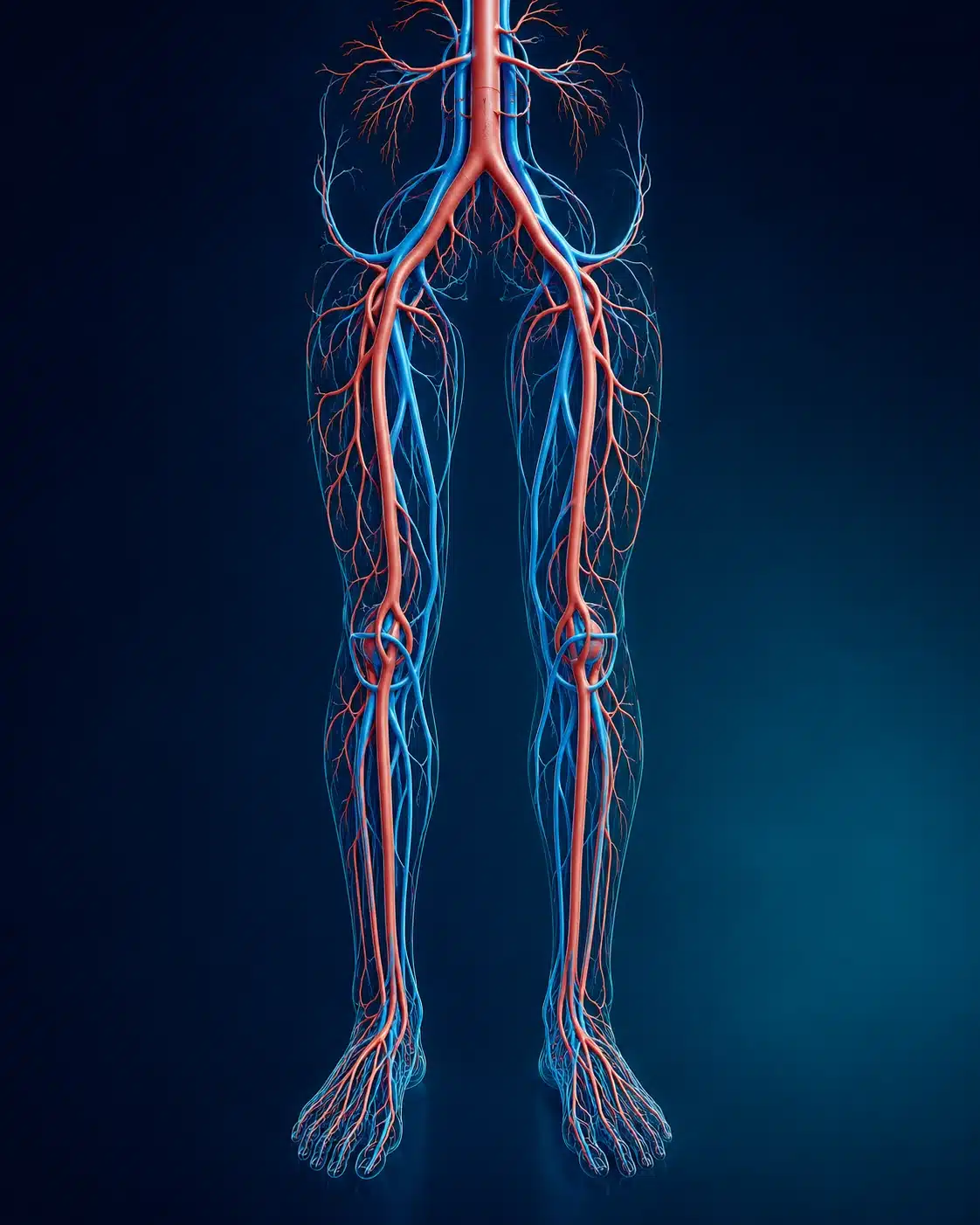
These medical visuals show the anatomy, treatment target and image-guided route involved in Bronchial Artery Embolisation . The actual procedure plan is based on the patient’s scans, symptoms and overall health.
Signs and symptoms
Evaluation & planning
A precise, image-guided pathway
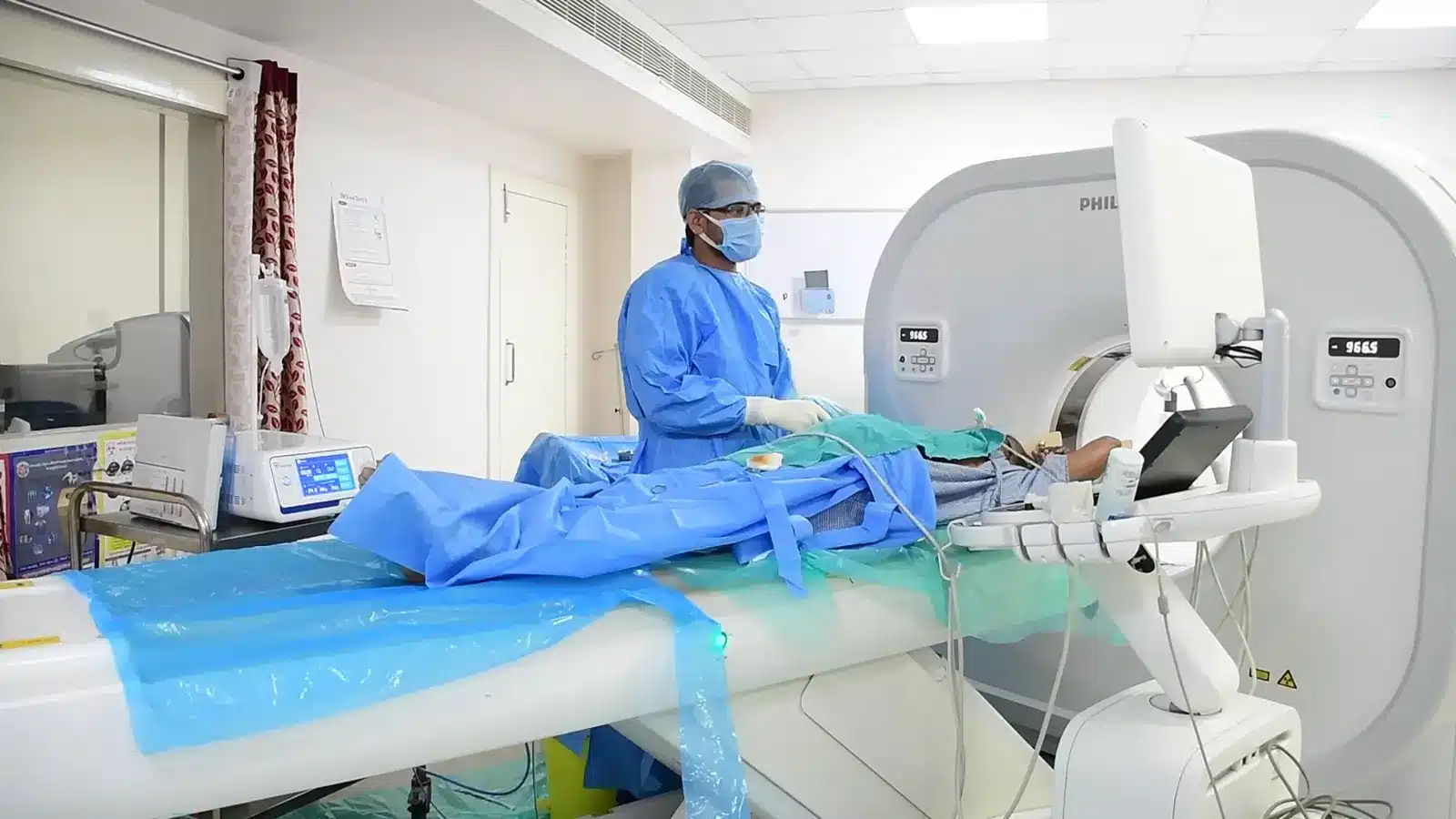
During Bronchial Artery Embolisation , imaging helps the interventional radiologist follow the anatomy, position the catheter or needle and confirm the treatment target. The exact technique and devices are chosen for the patient’s diagnosis and scan findings.
An artery is accessed through a small puncture.
Angiography maps abnormal bronchial vessels.
A microcatheter is positioned beyond protected branches.
Particles or coils are delivered to control bleeding.

Recovery and follow-up
Observation depends on the amount of bleeding and underlying illness. Mild chest discomfort or swallowing discomfort can occur. Recurrent bleeding, breathing difficulty or weakness needs emergency care.
After bronchial artery embolisation, the team monitors breathing, blood loss and the arterial puncture site. Temporary chest or swallowing discomfort can occur. Risks include access-site bleeding, contrast reaction and unintended embolisation, including rare serious neurological injury. New weakness or numbness, recurrent bleeding or breathing difficulty needs urgent medical attention. See the doctor’s vascular and interventional radiology profile. Bleeding from the digestive tract is a different problem, covered in the GI bleeding guide.

Meet Dr. M. Bhavani Shankar Manam
MBBS, MD, DNB (Radio Diagnosis), FIVR
Consultant Vascular & Interventional Radiologist with experience across vascular, non-vascular and oncological image-guided procedures and more than 3,000 cases. Every treatment plan begins with a review of symptoms, reports and imaging, followed by a clear discussion of suitable options.
Frequently asked questions
These answers are general and cannot determine your suitability without clinical review.
It controls bleeding vessels; infection, tumour or other underlying disease still needs treatment.
Yes. Rebleeding may occur and can require repeat embolisation or another treatment.
CT often helps identify the likely source and plan vessels, but emergency decisions depend on stability.
You may also want to understand
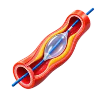
Peripheral Angioplasty
Peripheral angioplasty is a minimally invasive treatment used in selected patients to reopen narrowed or blocked arteries, mo …
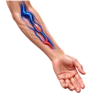
Fistuloplasty
Fistuloplasty is an angioplasty performed to widen a narrowed dialysis fistula or graft. By improving blood flow through the …
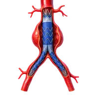
Aorta Stenting
Aortic stent-graft repair is a minimally invasive treatment for selected aortic aneurysms, dissections and injuries. A fabric …