Image-Guided Biopsy, Drainage & Access in Vijayawada
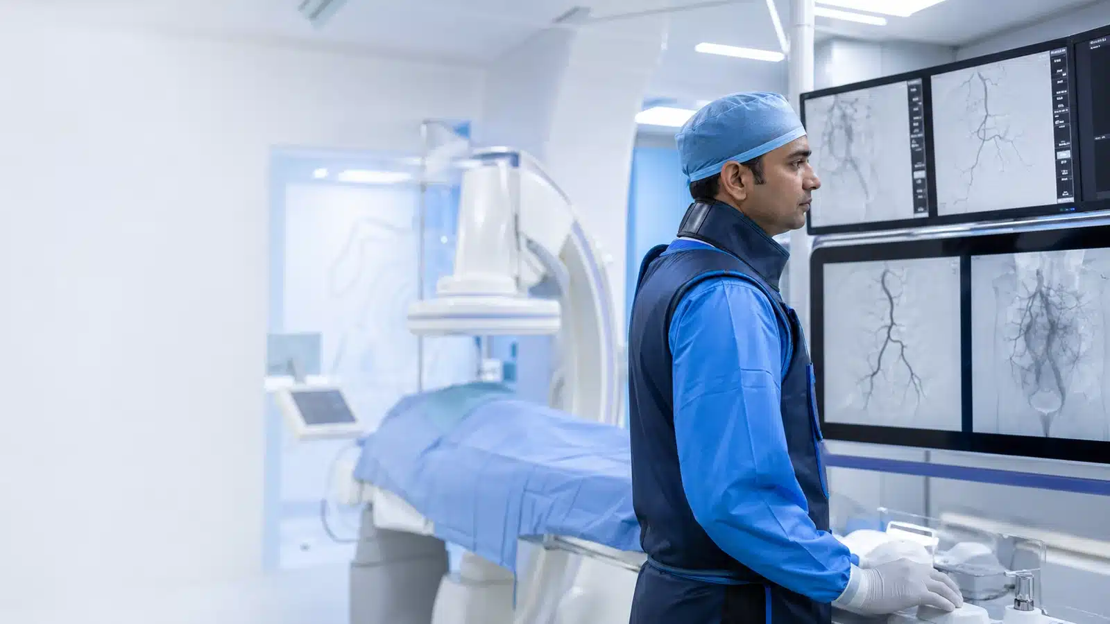
Image-guided biopsy in Vijayawada uses ultrasound or CT to direct a needle to a selected tissue target for laboratory examination. Related procedures include image-guided abscess drainage and catheter placement for blocked fluid pathways or medical access. A vascular and interventional radiologist reviews the referral, scans and blood tests to choose an appropriate route and explain the purpose of the procedure.
Understanding Biopsy & Drainage
Image guidance helps choose a safe path to a target while avoiding important vessels and organs. Biopsy provides tissue for pathology; drainage removes infected or obstructed fluid; access procedures create a route for medicines, nutrition, dialysis or urinary and biliary drainage.
An image guided biopsy in Vijayawada is planned from recent scans and blood tests. The type of anaesthetic, needle and observation time depend on the organ and procedure. Blood thinners may need specific instructions and should never be stopped without medical advice.
A biopsy obtains tissue for diagnosis; drainage removes a collection or relieves an obstruction. They are different procedures with different preparation and aftercare. Bring previous scan images, referral notes, blood tests and your medicine list. Tell the team about allergies, blood thinners and any possibility of pregnancy. The organ being sampled determines the needle type, local anaesthetic or sedation and observation period. A sample may occasionally be insufficient and require repeat biopsy. Laboratory reporting time depends on the tests requested.
Understanding the condition and treatment pathway
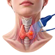
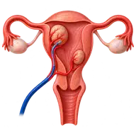
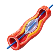
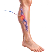
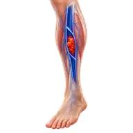
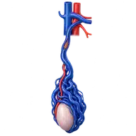
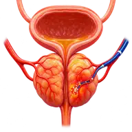
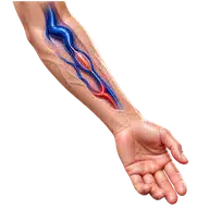
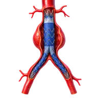
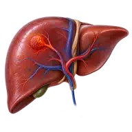
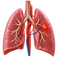
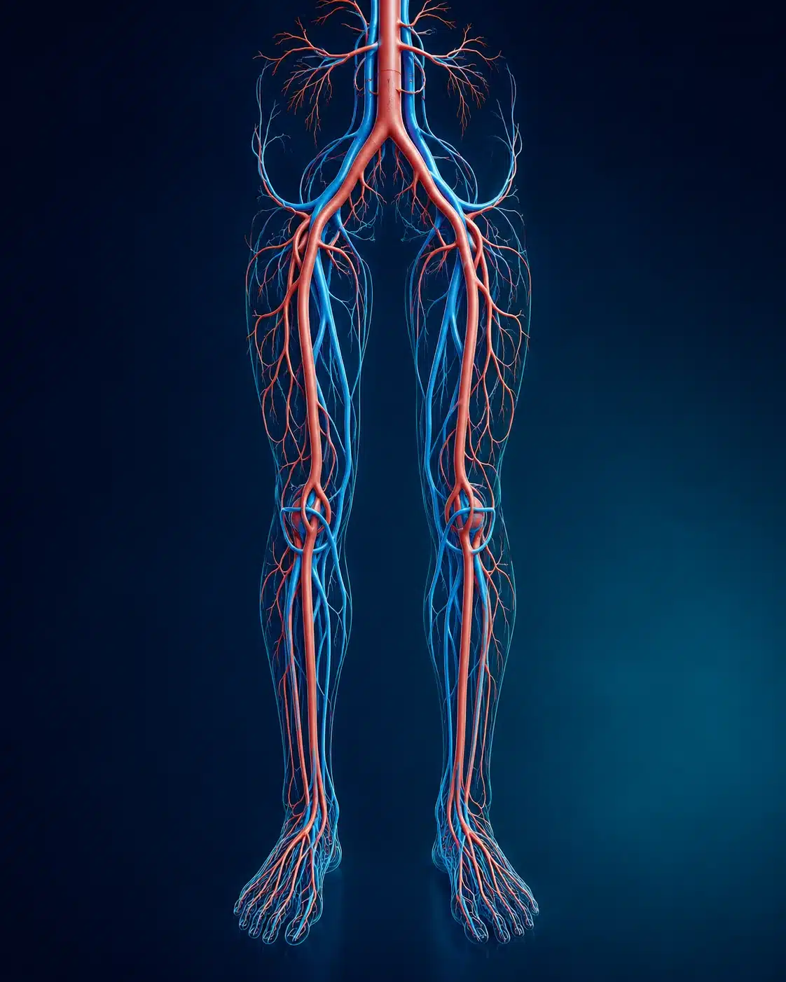
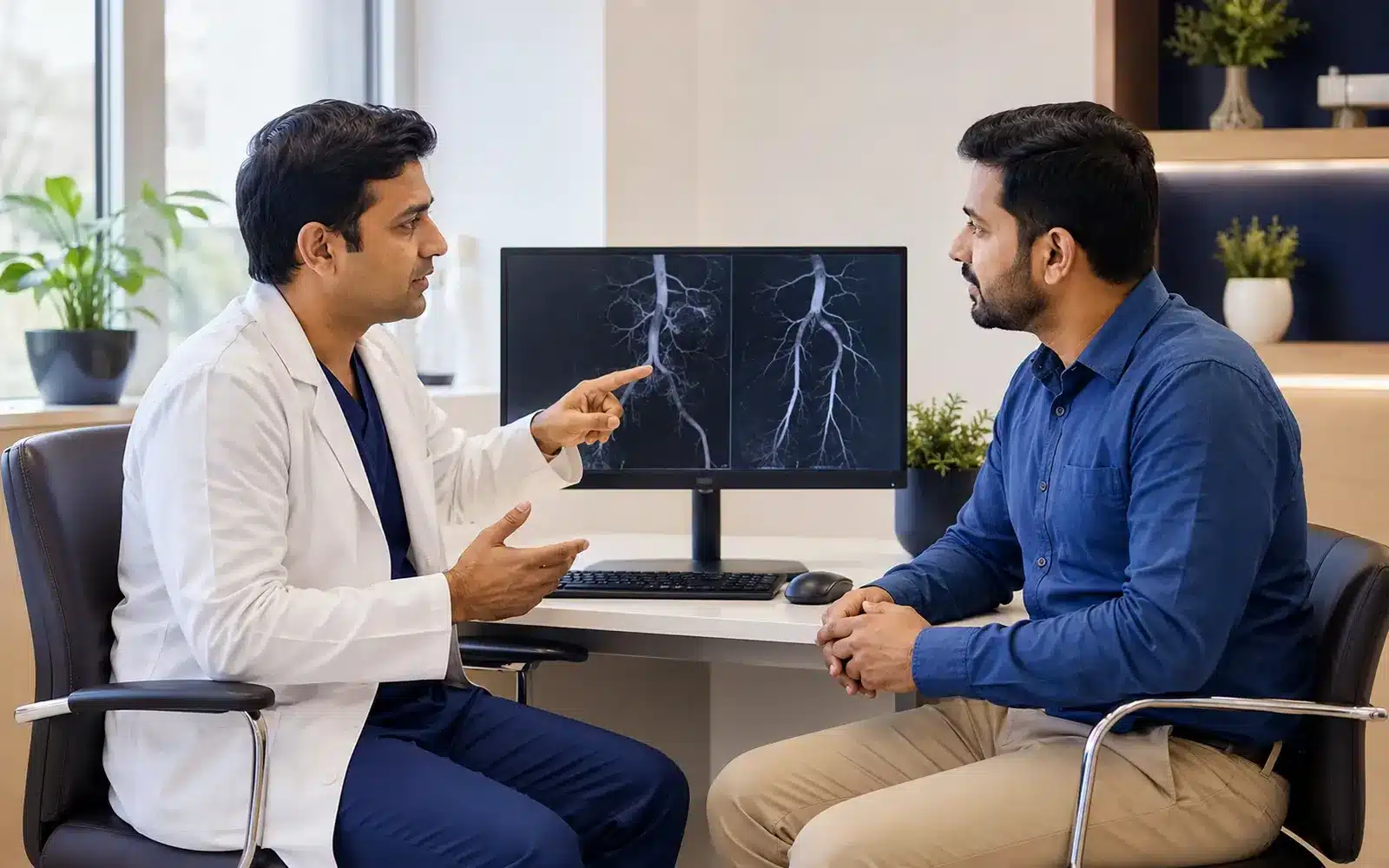
These medical visuals show the anatomy, treatment target and image-guided route involved in Biopsy & Drainage . The actual procedure plan is based on the patient’s scans, symptoms and overall health.
Signs and symptoms
Evaluation & planning
A precise, image-guided pathway
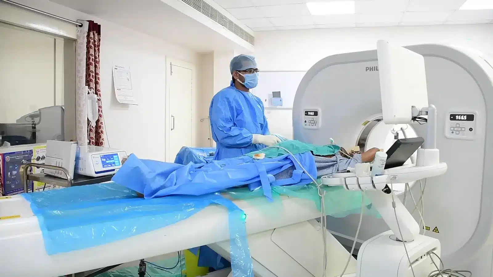
During Biopsy & Drainage , imaging helps the interventional radiologist follow the anatomy, position the catheter or needle and confirm the treatment target. The exact technique and devices are chosen for the patient’s diagnosis and scan findings.
Imaging selects the safest access path.
Local anaesthetic and sedation are used as appropriate.
A needle, catheter or port is placed precisely.
Samples or fluid are sent for laboratory analysis when needed.
Recovery and follow-up
Observation and restrictions depend on the organ and procedure. Fever, increasing pain, breathlessness, bleeding or catheter problems should be reported urgently using the instructions provided.
After image-guided biopsy or drainage, follow the organ-specific instructions on rest, dressings and return to activity. Bleeding and infection are possible risks; a lung biopsy also carries a risk of air leaking around the lung. A drainage catheter needs instructions for care, output monitoring and planned review or removal. Do not flush or remove it without the team’s advice. Report breathlessness, heavy bleeding, fever or increasing pain promptly. See interventional oncology for treatment procedures, or request an assessment with the relevant reports.
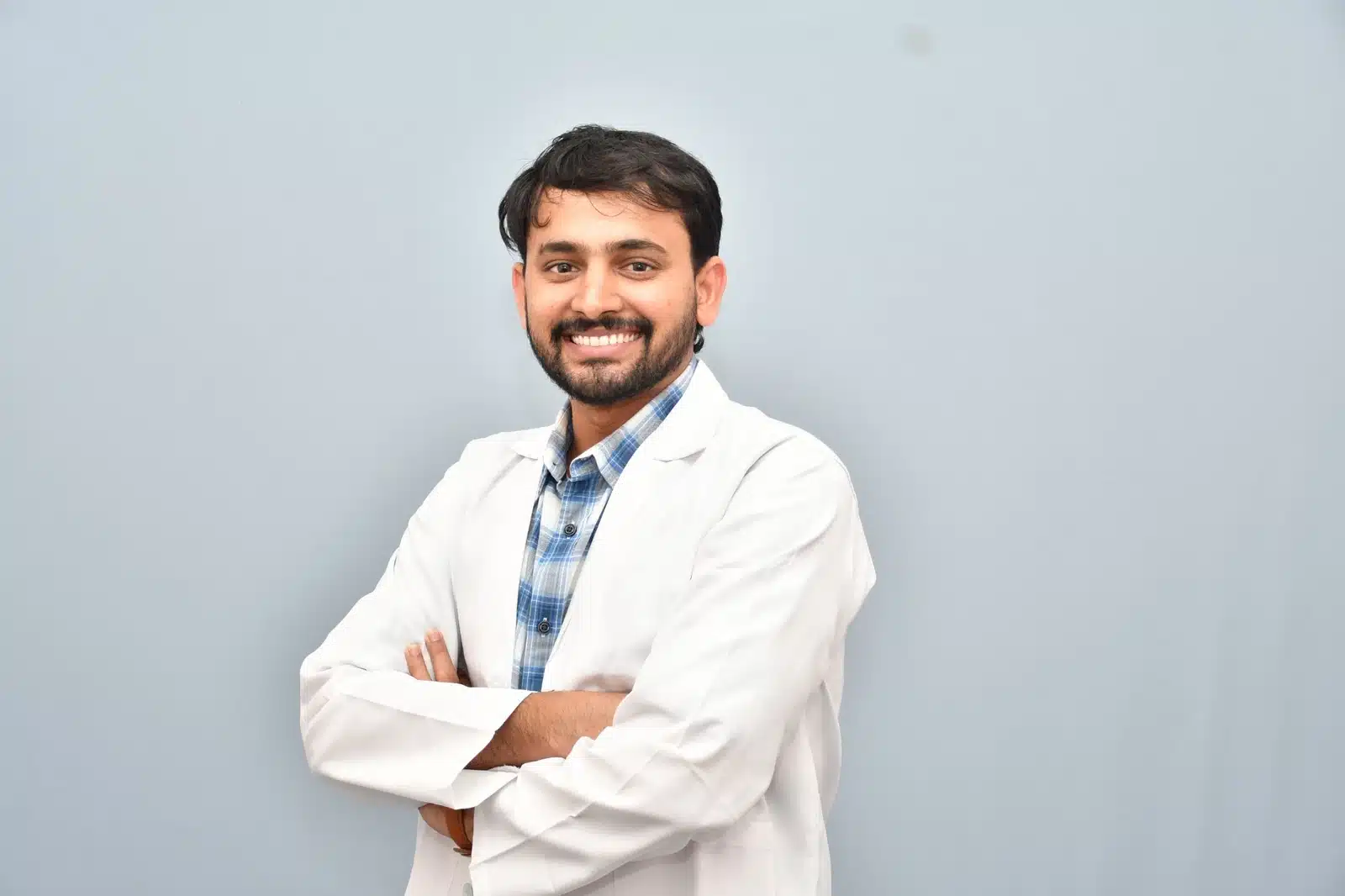
Meet Dr. M. Bhavani Shankar Manam
MBBS, MD, DNB (Radio Diagnosis), FIVR
Consultant Vascular & Interventional Radiologist with experience across vascular, non-vascular and oncological image-guided procedures and more than 3,000 cases. Every treatment plan begins with a review of symptoms, reports and imaging, followed by a clear discussion of suitable options.
Frequently asked questions
These answers are general and cannot determine your suitability without clinical review.
Modern image-guided biopsy is widely used; the route and technique are planned to minimise risks.
No. Some catheters remain for days or weeks; others are removed after a single aspiration.
Pathology timing varies by sample and any special tests required.