Deep Vein Thrombosis Evaluation & Treatment in Vijayawada
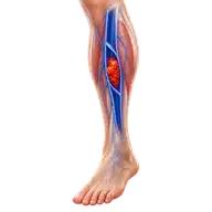
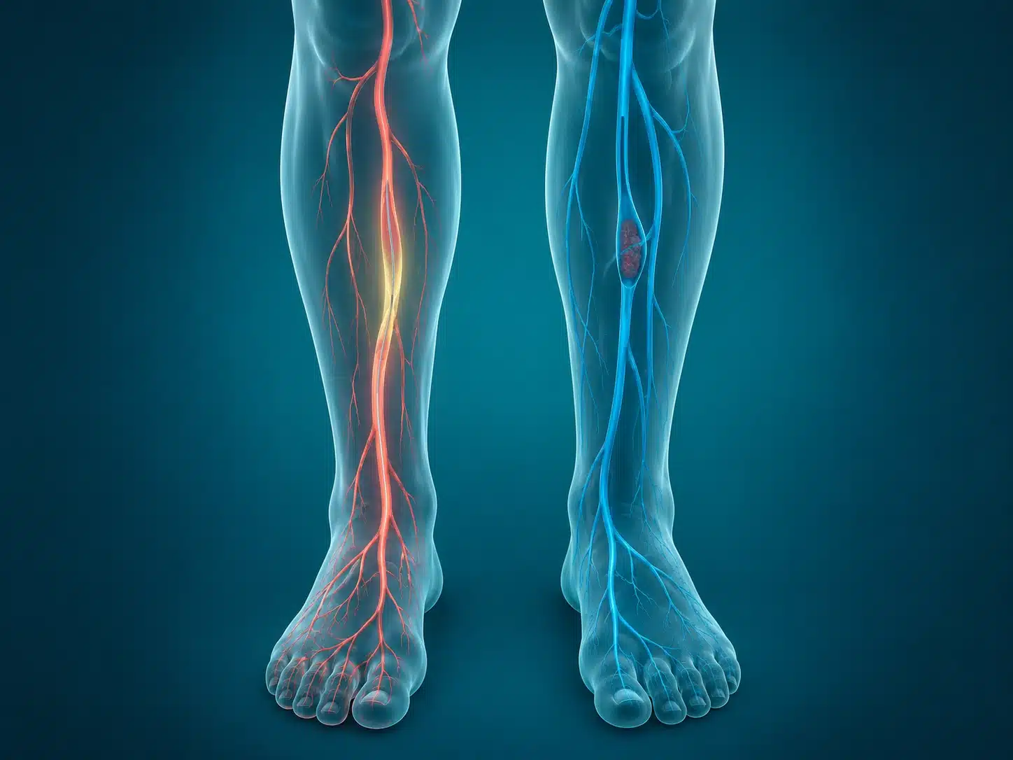
Deep vein thrombosis (DVT) is a blood clot in a deep vein, usually in the leg. It requires timely diagnosis because part of the clot can travel to the lungs. Most patients need anticoagulant medicine; selected extensive or limb-threatening clots may need catheter-based treatment.
DVT can cause new one-sided swelling, pain, warmth, heaviness or colour change, although some clots produce few symptoms. Risk can rise after major surgery, prolonged immobility, pregnancy, cancer, serious illness or a previous clot. Sudden breathlessness, chest pain, fainting or coughing blood is an emergency.
DVT treatment in Vijayawada starts with clinical assessment and Doppler ultrasound. Blood thinners are the foundation for most patients. In carefully selected patients with a large recent clot, severe symptoms or threatened circulation, catheter-directed thrombolysis or mechanical thrombectomy may be considered after weighing bleeding risk.
The long-term plan can include anticoagulation duration, compression when appropriate, activity guidance and evaluation for an underlying cause. Follow-up also watches for post-thrombotic syndrome, which may cause persistent swelling, heaviness, skin changes or ulcers.
Understanding the condition and treatment pathway
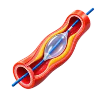
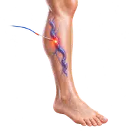
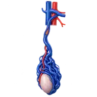
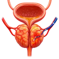
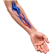
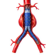
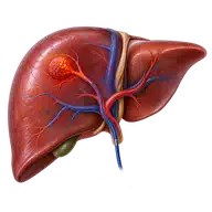
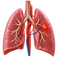
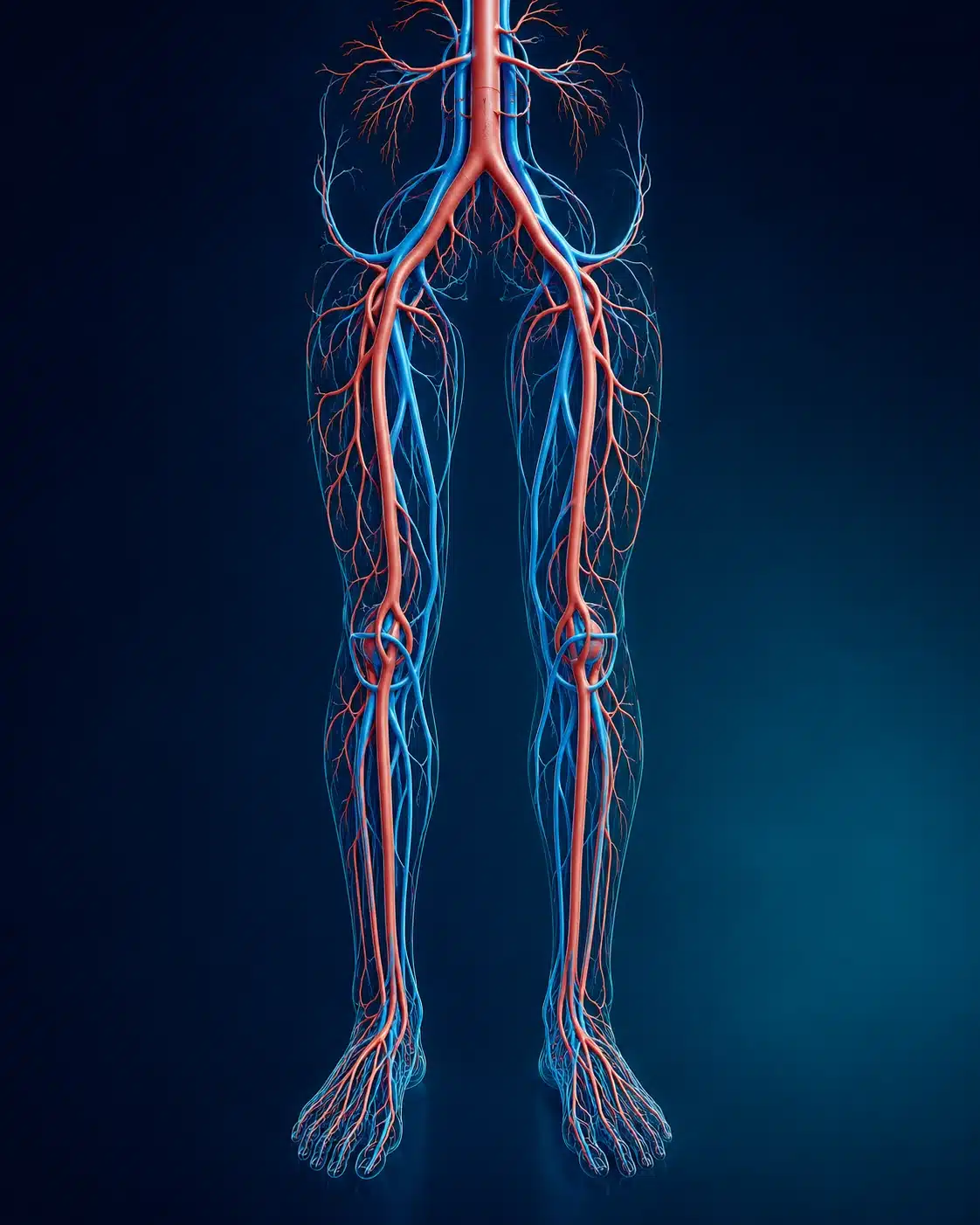
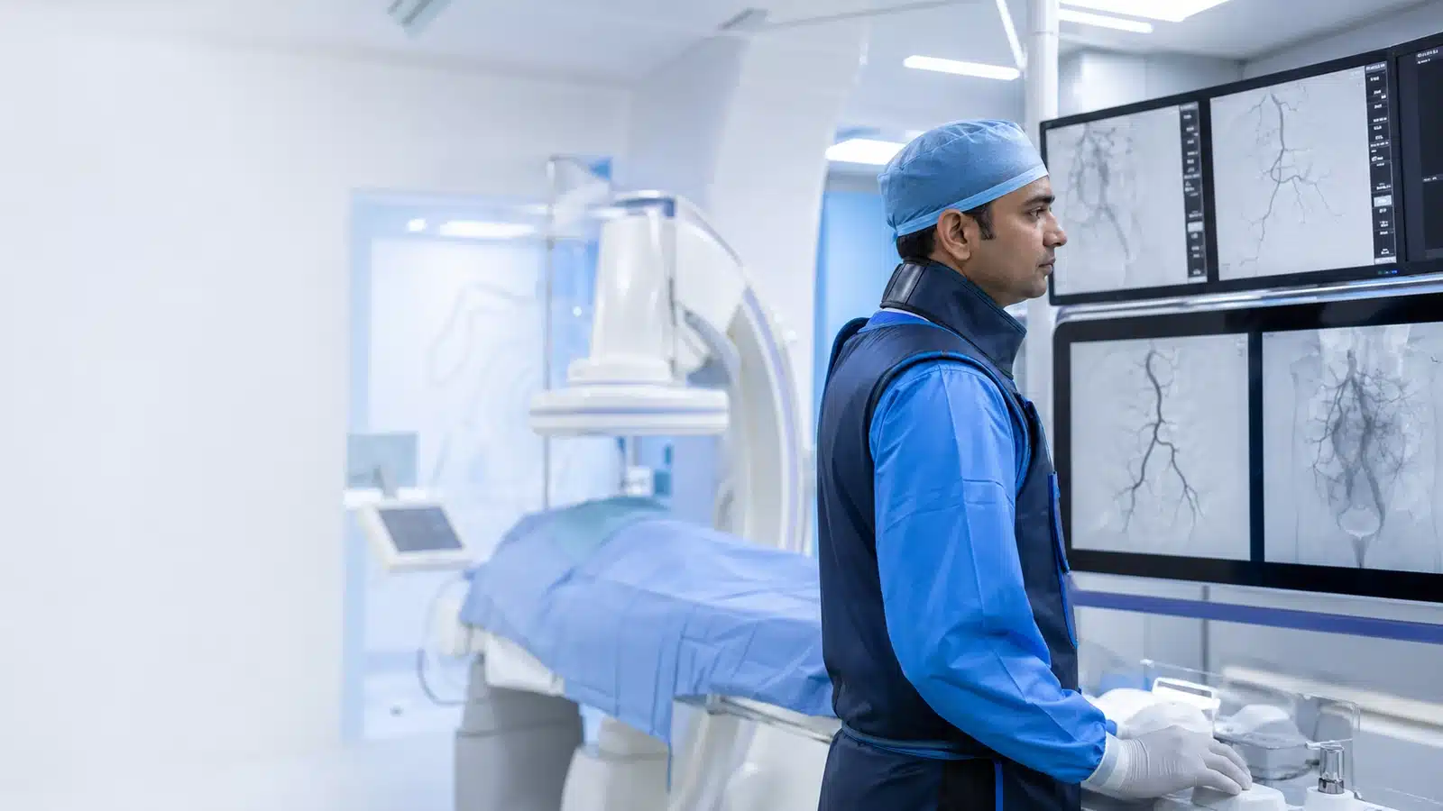
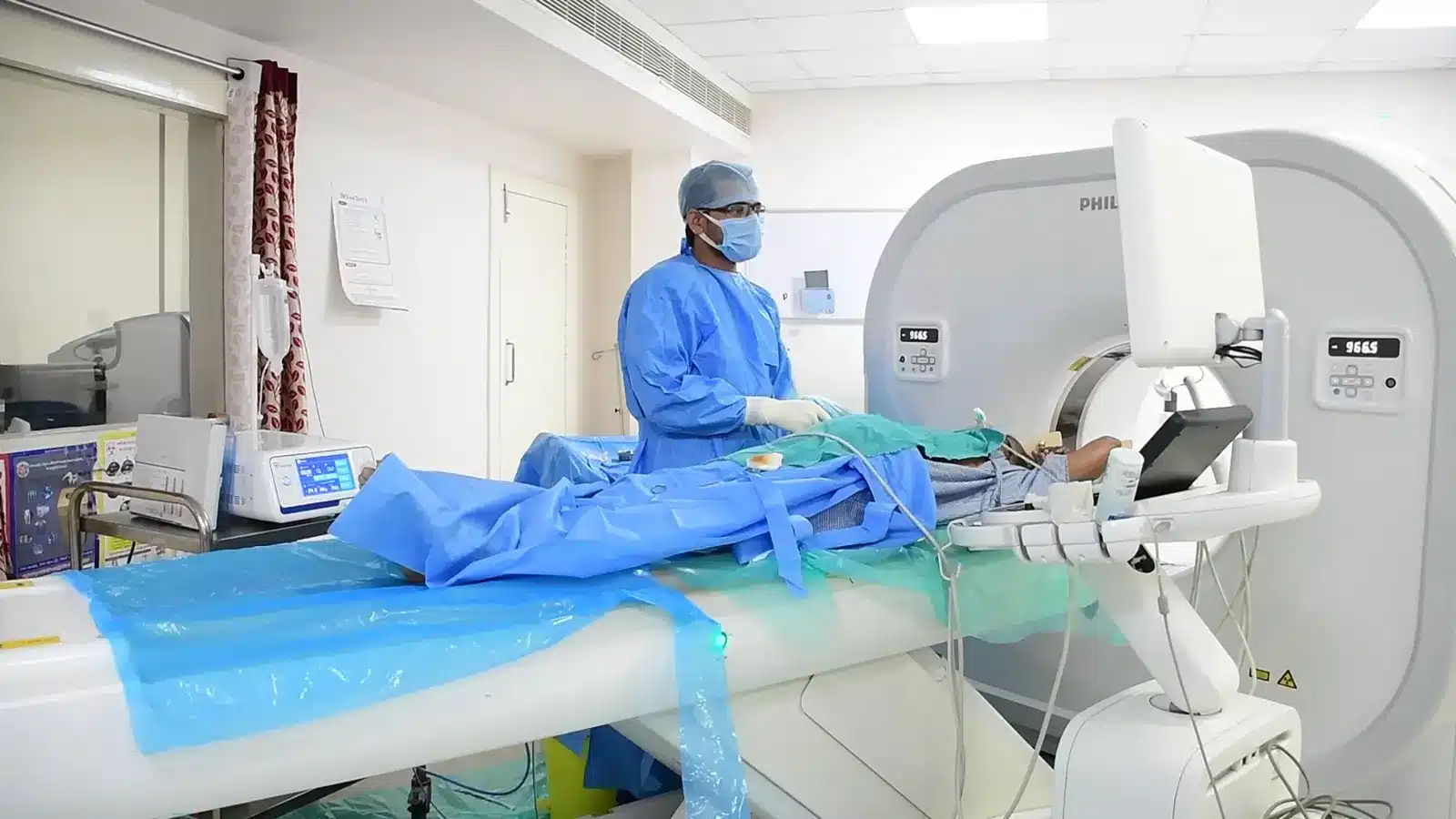
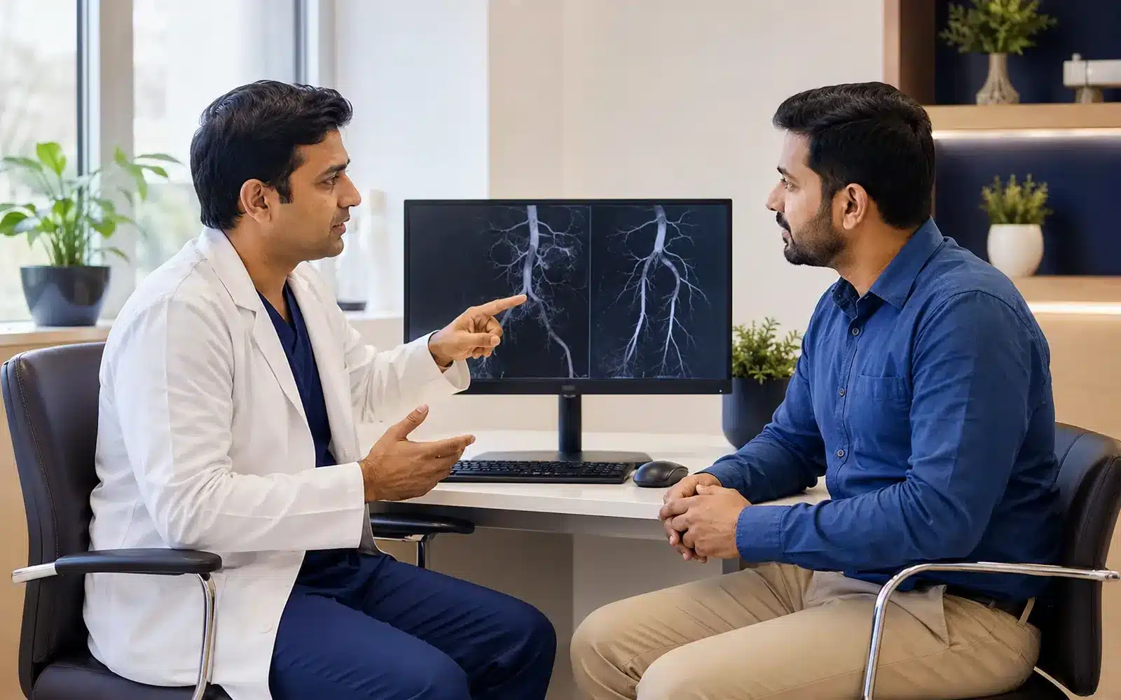
These medical visuals show the anatomy, treatment target and image-guided route involved in Deep Vein Thrombosis. The actual procedure plan is based on the patient’s scans, symptoms and overall health.
Signs and symptoms
✓
New one-sided leg swelling
✓
Calf or thigh pain and warmth
✓
Skin colour change or heaviness
✓
Breathlessness or chest pain — emergency
Evaluation & planning
✓
Clinical risk assessment
✓
Venous Doppler ultrasound
✓
Blood and kidney function tests
✓
Review of bleeding risk and provoking factors
A precise, image-guided pathway
During Deep Vein Thrombosis, imaging helps the interventional radiologist follow the anatomy, position the catheter or needle and confirm the treatment target. The exact technique and devices are chosen for the patient’s diagnosis and scan findings.
01
Most patients begin anticoagulation when appropriate.
02
Imaging defines clot location and extent.
03
Selected severe cases may receive catheter-based clot removal or clot-dissolving treatment.
04
A follow-up plan helps reduce recurrence and chronic symptoms.
Recovery and follow-up
Activity, compression and travel advice differ by patient. Anticoagulants must be taken exactly as prescribed. New bleeding, worsening swelling, breathlessness or chest pain requires immediate medical attention.
Before discharge, the team explains medicines, activity, wound or access-site care, expected symptoms and the warning signs that should prompt urgent review. Follow-up timing depends on the condition and may include Doppler, CT, MRI, blood tests or consultation with another specialist.
Consultant Vascular & Interventional Radiologist with experience across vascular, non-vascular and oncological image-guided procedures and more than 3,000 cases. Every treatment plan begins with a review of symptoms, reports and imaging, followed by a clear discussion of suitable options.