Image-Guided Thyroid Nodule Ablation in Vijayawada
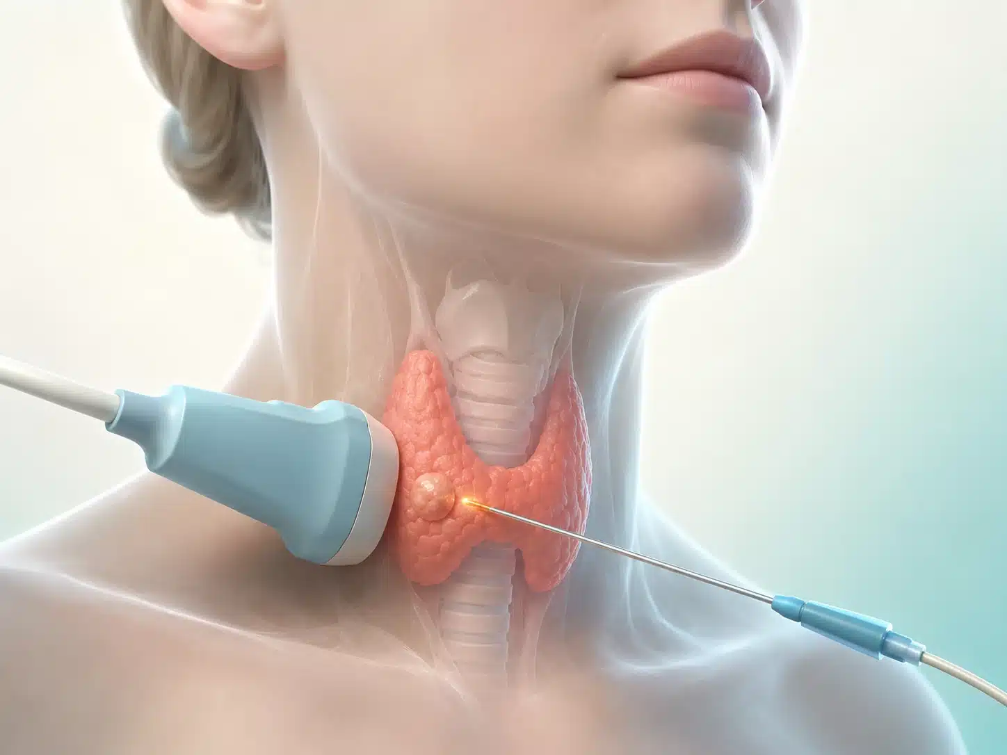
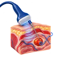
Radiofrequency ablation (RFA) can shrink selected benign thyroid nodules that cause pressure, swallowing discomfort or a visible neck swelling. A thin needle electrode is placed into the nodule under continuous ultrasound guidance while the patient remains awake.
Most thyroid nodules do not need ablation. The first steps are thyroid blood tests, high-quality ultrasound and biopsy when indicated. RFA is generally considered for nodules confirmed as benign and causing meaningful symptoms or cosmetic concern.
During thyroid nodule ablation in Vijayawada, local anaesthetic is used and the electrode is moved through small zones of the nodule to deliver controlled heat. Ultrasound helps protect the voice nerve, blood vessels, trachea and normal thyroid tissue.
The nodule shrinks over several months; it does not disappear immediately. Surgery, observation, medication or other ablation methods may be more appropriate depending on diagnosis, size, location and thyroid function.
Understanding the condition and treatment pathway
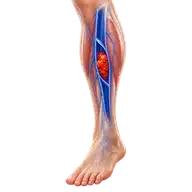
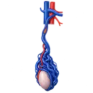
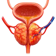
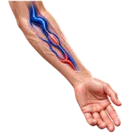
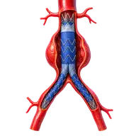
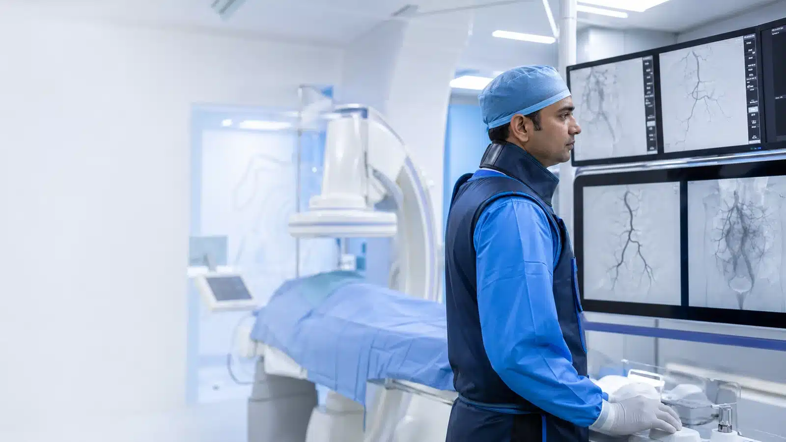
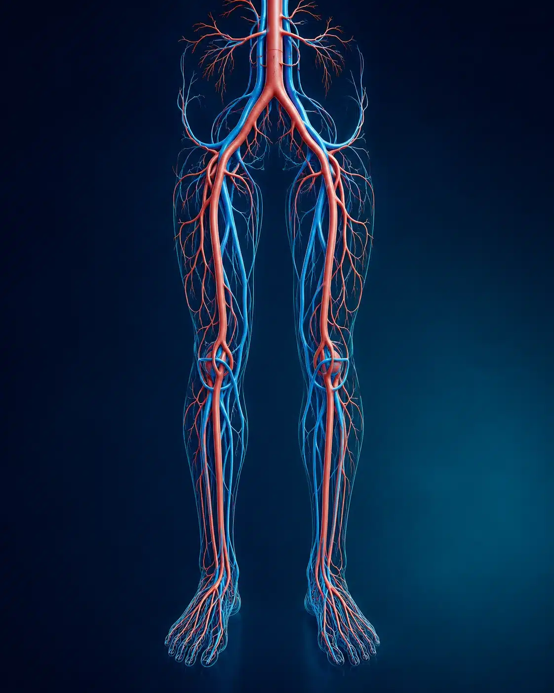
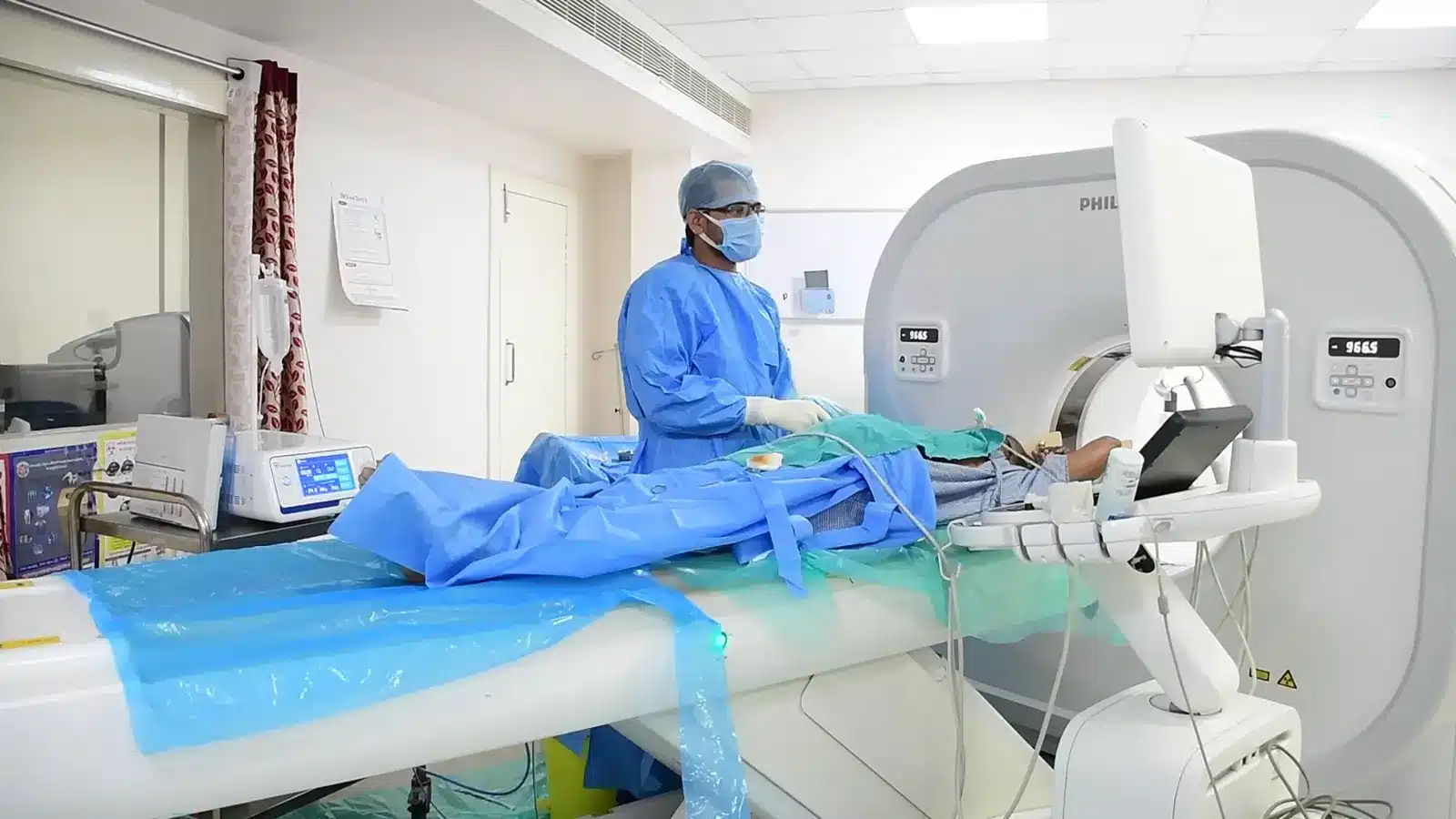
These medical visuals show the anatomy, treatment target and image-guided route involved in Thyroid Nodule Ablation. The actual procedure plan is based on the patient’s scans, symptoms and overall health.
Signs and symptoms
✓
Visible or enlarging neck lump
✓
Pressure or swallowing discomfort
✓
Benign nodule causing local symptoms
✓
Cosmetic concern after appropriate evaluation
Evaluation & planning
✓
Detailed thyroid ultrasound
✓
Thyroid function blood tests
✓
Benign biopsy confirmation when indicated
✓
Voice and symptom assessment
A precise, image-guided pathway
During Thyroid Nodule Ablation, imaging helps the interventional radiologist follow the anatomy, position the catheter or needle and confirm the treatment target. The exact technique and devices are chosen for the patient’s diagnosis and scan findings.
01
Ultrasound maps the nodule and nearby structures.
02
Local anaesthetic is applied.
03
A fine RFA electrode treats the nodule in small controlled zones.
04
Ultrasound checks the treated area before discharge.
Recovery and follow-up
Temporary neck discomfort, swelling or voice change can occur. Patients receive advice about eating, medicines and warning signs. Ultrasound follow-up measures volume reduction and checks for regrowth.
Before discharge, the team explains medicines, activity, wound or access-site care, expected symptoms and the warning signs that should prompt urgent review. Follow-up timing depends on the condition and may include Doppler, CT, MRI, blood tests or consultation with another specialist.
Consultant Vascular & Interventional Radiologist with experience across vascular, non-vascular and oncological image-guided procedures and more than 3,000 cases. Every treatment plan begins with a review of symptoms, reports and imaging, followed by a clear discussion of suitable options.