Bronchial Artery Embolisation for Coughing Blood in Vijayawada
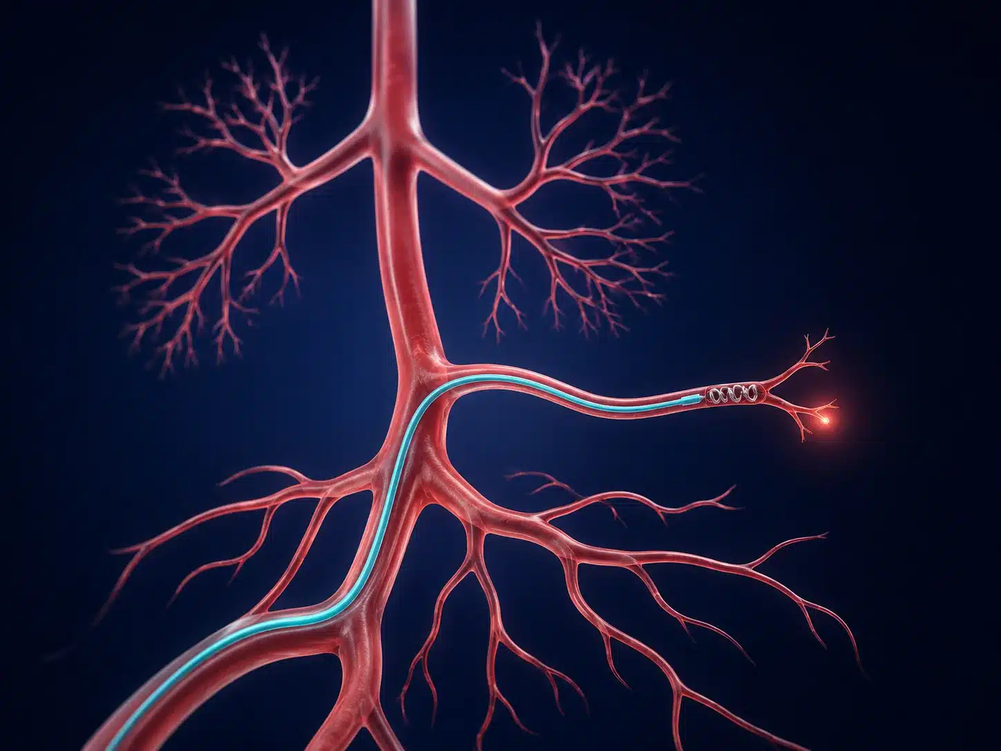
Bronchial artery embolisation (BAE) is an image-guided emergency or planned treatment used to control significant or recurrent coughing of blood. A microcatheter is guided into abnormal bronchial or other systemic arteries and embolic material is delivered to stop the bleeding source.
Coughing blood, called haemoptysis, can occur with infection, bronchiectasis, tuberculosis-related lung damage, tumours, vascular abnormalities and other conditions. Even a smaller amount can become urgent when bleeding continues, breathing is affected or the patient is medically fragile.
Bronchial artery embolisation in Vijayawada is planned using CT and angiography. The interventional radiologist maps arteries supplying the abnormal lung area while carefully identifying branches that must be protected. The goal is to control bleeding; the underlying lung disease still requires treatment.
BAE can be lifesaving, but rebleeding can occur, especially when the underlying condition progresses or additional vessels develop. Respiratory physicians, critical-care teams and thoracic surgeons may be involved depending on severity.
Understanding the condition and treatment pathway
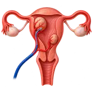
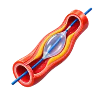
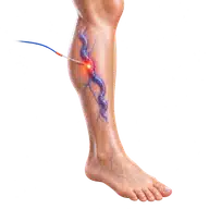
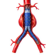
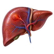
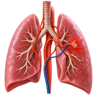
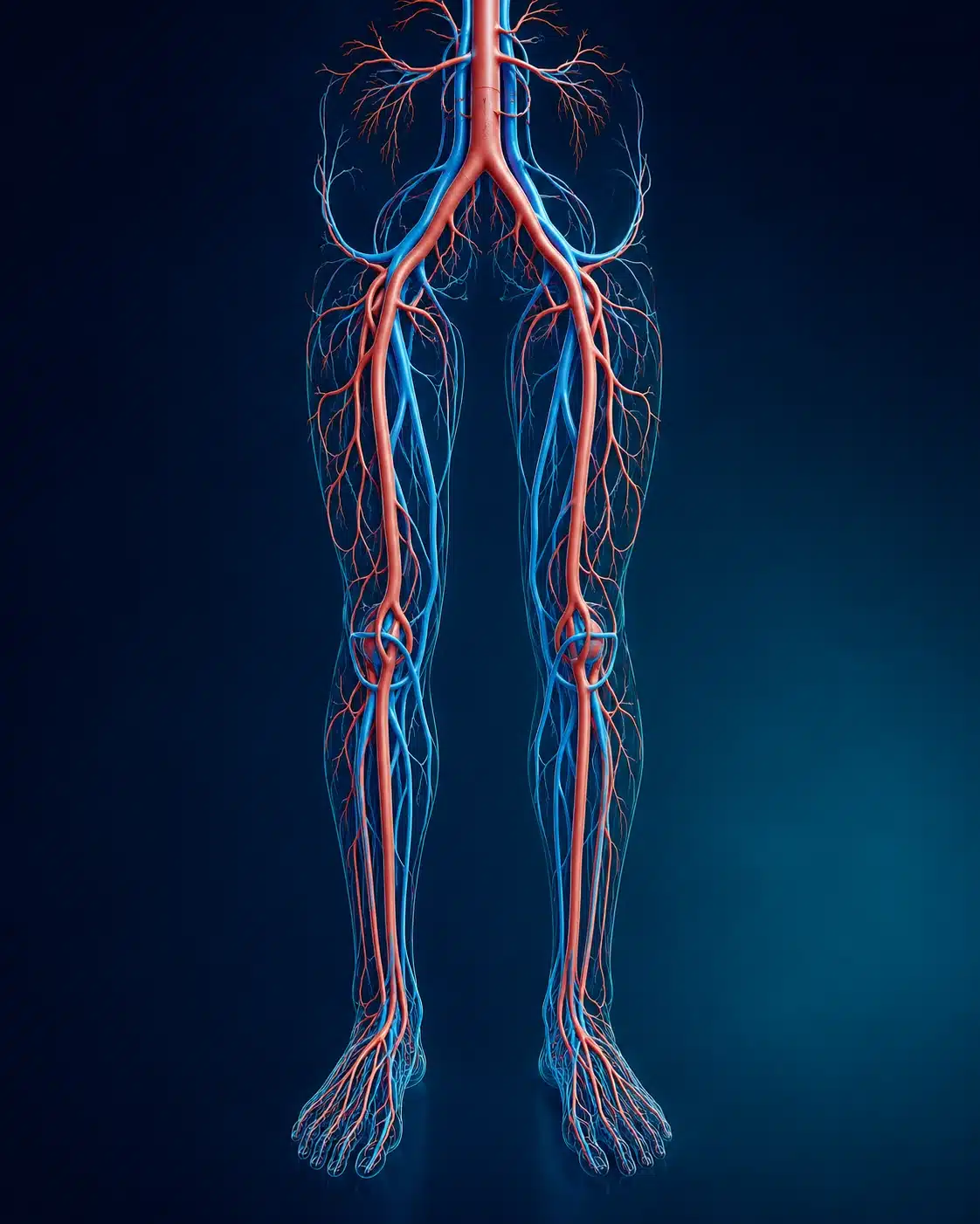
These medical visuals show the anatomy, treatment target and image-guided route involved in Bronchial Artery Embolisation. The actual procedure plan is based on the patient’s scans, symptoms and overall health.
Signs and symptoms
✓
Coughing bright-red or blood-streaked sputum
✓
Repeated episodes of haemoptysis
✓
Breathlessness, dizziness or weakness
✓
Known bronchiectasis, TB damage or lung lesion
Evaluation & planning
✓
Airway and circulation assessment
✓
CT chest or CT angiography
✓
Blood count and clotting tests
✓
Respiratory and infection review
A precise, image-guided pathway
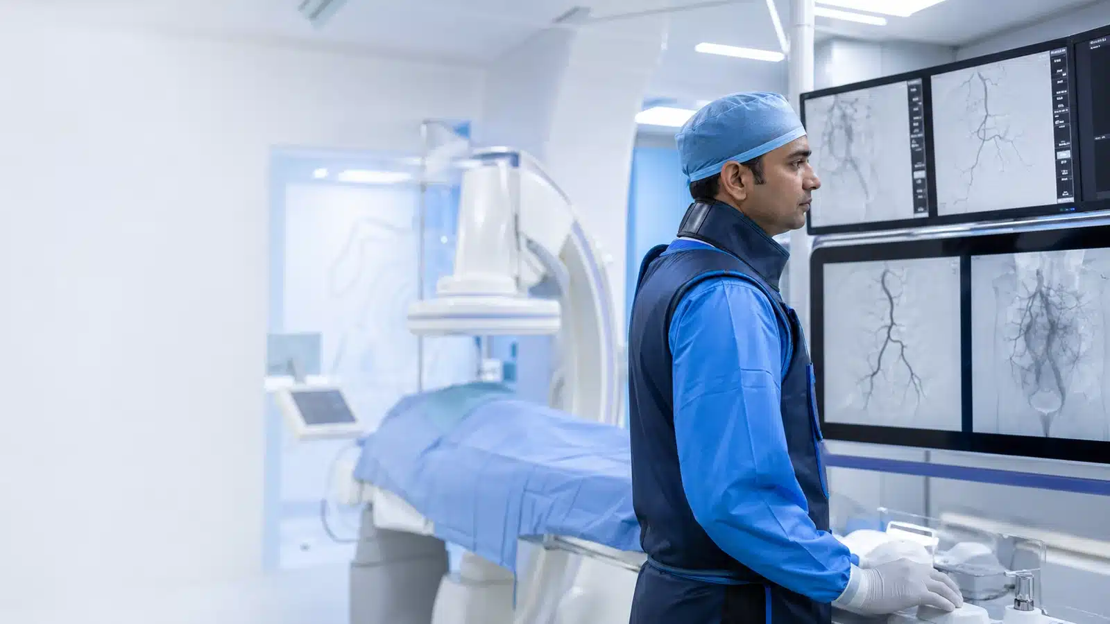
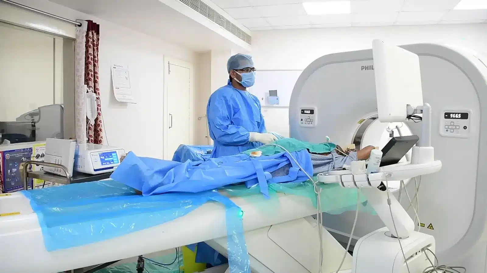
During Bronchial Artery Embolisation, imaging helps the interventional radiologist follow the anatomy, position the catheter or needle and confirm the treatment target. The exact technique and devices are chosen for the patient’s diagnosis and scan findings.
01
An artery is accessed through a small puncture.
02
Angiography maps abnormal bronchial vessels.
03
A microcatheter is positioned beyond protected branches.
04
Particles or coils are delivered to control bleeding.
Recovery and follow-up
Observation depends on the amount of bleeding and underlying illness. Mild chest discomfort or swallowing discomfort can occur. Recurrent bleeding, breathing difficulty or weakness needs emergency care.
Before discharge, the team explains medicines, activity, wound or access-site care, expected symptoms and the warning signs that should prompt urgent review. Follow-up timing depends on the condition and may include Doppler, CT, MRI, blood tests or consultation with another specialist.
Consultant Vascular & Interventional Radiologist with experience across vascular, non-vascular and oncological image-guided procedures and more than 3,000 cases. Every treatment plan begins with a review of symptoms, reports and imaging, followed by a clear discussion of suitable options.