Aortic Stent Graft Treatment in Vijayawada
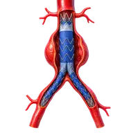
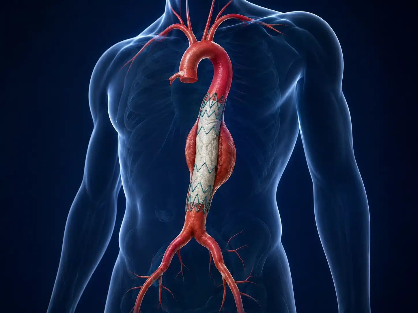
Aortic stent-graft repair is a minimally invasive treatment for selected aortic aneurysms, dissections and injuries. A fabric-covered stent is guided through the arteries and positioned inside the aorta to exclude the diseased segment from blood pressure and flow.
Understanding Aorta Stenting
The aorta is the body’s main artery. An aneurysm is an abnormal enlargement that may rupture; a dissection is a tear within the aortic wall. These conditions require expert assessment because urgency and treatment differ according to location, size, symptoms, growth and anatomy.
Aorta stenting in Vijayawada may involve endovascular aneurysm repair (EVAR) in the abdomen or thoracic endovascular aortic repair (TEVAR) in the chest. Detailed CT angiography is used to measure landing zones, branch vessels and access arteries. Some complex cases need open surgery or a hybrid approach.
After stent-graft treatment, lifelong surveillance imaging is important. Follow-up checks the graft position, aneurysm sac and possible endoleak. Blood-pressure control, smoking cessation and cardiovascular prevention continue after the procedure.
Understanding the condition and treatment pathway
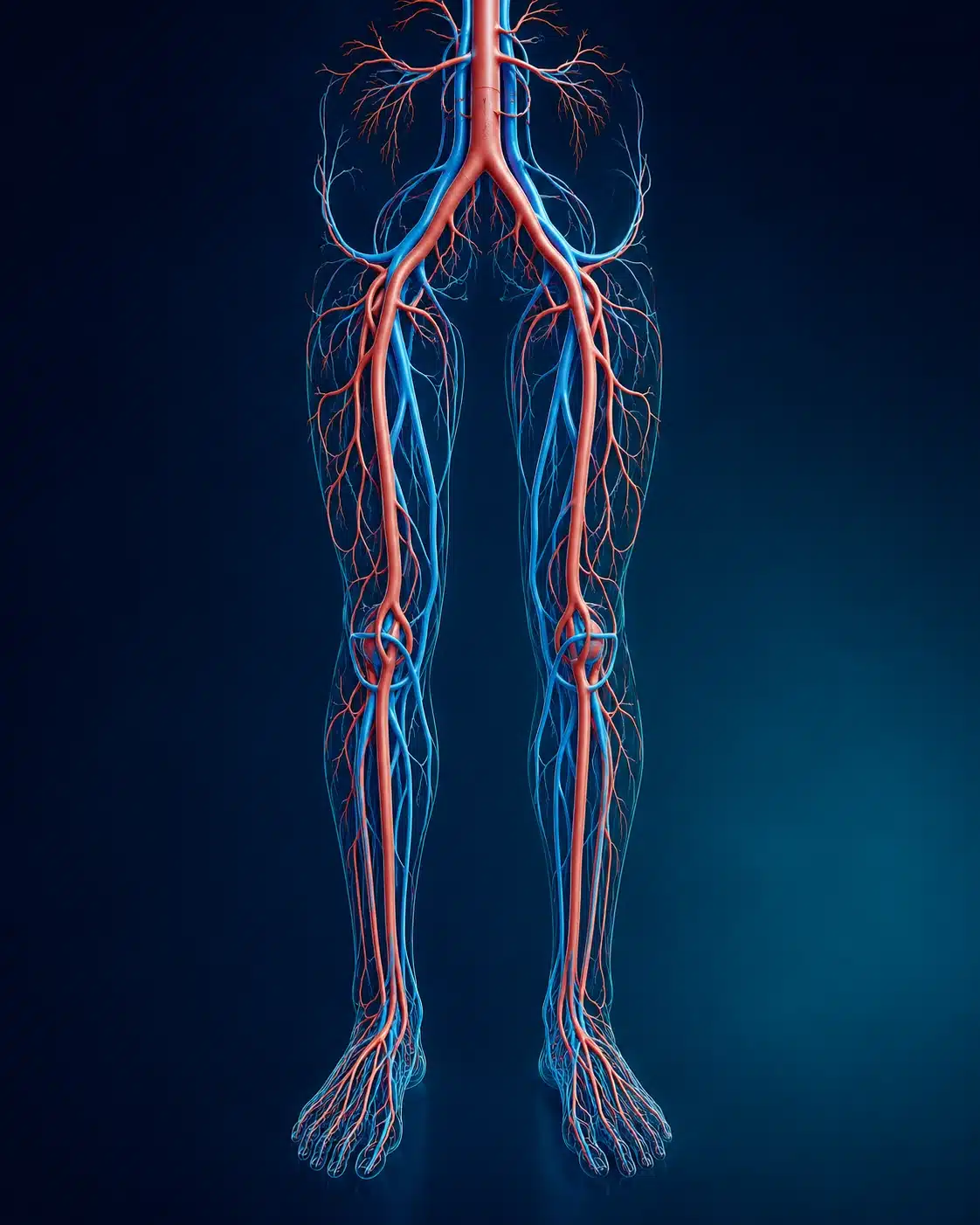
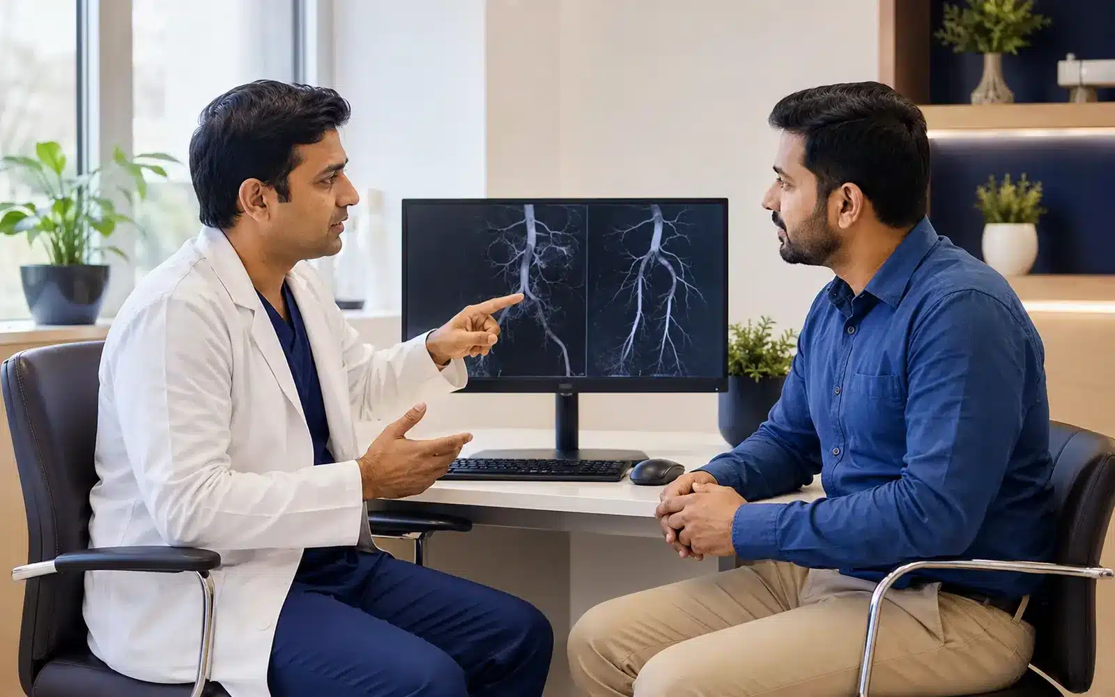
These medical visuals show the anatomy, treatment target and image-guided route involved in Aorta Stenting . The actual procedure plan is based on the patient’s scans, symptoms and overall health.
Signs and symptoms
Evaluation & planning
A precise, image-guided pathway
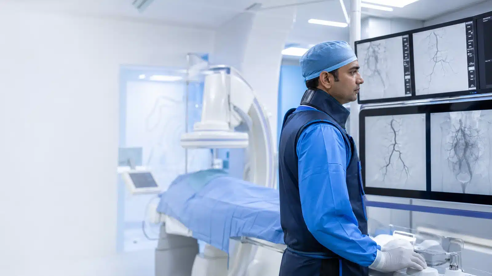
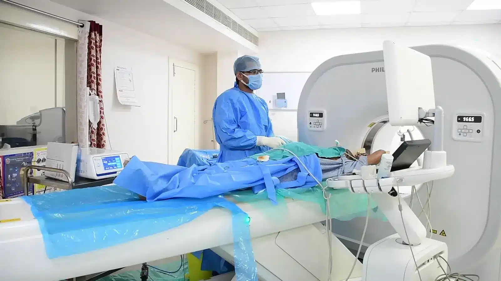
During Aorta Stenting , imaging helps the interventional radiologist follow the anatomy, position the catheter or needle and confirm the treatment target. The exact technique and devices are chosen for the patient’s diagnosis and scan findings.
Access arteries are prepared, usually in the groins.
Guidewires position the stent graft under X-ray guidance.
The graft is deployed across the diseased segment.
Angiography checks sealing and branch circulation.
Recovery and follow-up
Hospital monitoring depends on the aortic condition and procedure complexity. Follow-up CT or ultrasound is essential. New severe pain, fainting, weakness or circulation change needs emergency assessment.
Aortic stent-graft treatment requires detailed CT planning and discussion of open repair, endovascular repair or surveillance. Risks include access-vessel injury, bleeding, kidney injury from contrast, graft complications and reduced blood flow to organs or limbs. Follow-up checks for an endoleak, where blood continues to enter the aneurysm outside the graft. Further treatment may be necessary even after an initially satisfactory repair. Sudden severe chest, back or abdominal pain, fainting or weakness requires emergency assessment. The vascular and interventional radiologist reviews the scans with the wider treatment team.
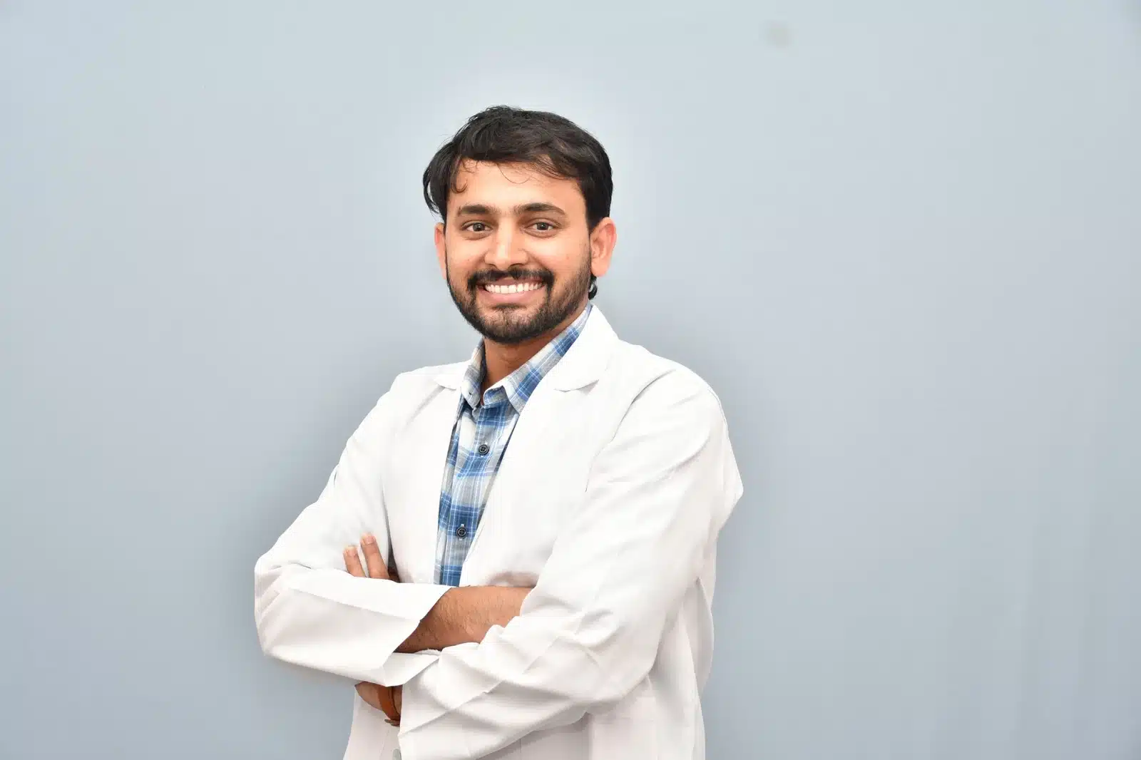
Meet Dr. M. Bhavani Shankar Manam
MBBS, MD, DNB (Radio Diagnosis), FIVR
Consultant Vascular & Interventional Radiologist with experience across vascular, non-vascular and oncological image-guided procedures and more than 3,000 cases. Every treatment plan begins with a review of symptoms, reports and imaging, followed by a clear discussion of suitable options.
Frequently asked questions
These answers are general and cannot determine your suitability without clinical review.
No. The aortic shape, branch vessels, access arteries, age and health determine suitability.
Imaging checks graft position, sealing and the aneurysm sac over time.
Yes. Open or hybrid repair may be safer or more durable for some anatomy.
You may also want to understand
Peripheral Angioplasty
Peripheral angioplasty is a minimally invasive treatment used in selected patients to reopen narrowed or blocked arteries, mo …
Fistuloplasty
Fistuloplasty is an angioplasty performed to widen a narrowed dialysis fistula or graft. By improving blood flow through the …
Bronchial Artery Embolisation
Bronchial artery embolisation (BAE) is an image-guided emergency or planned treatment used to control significant or recurren …