Symptomatic uterine fibroids: the reported case
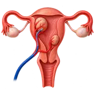
A 35-year-old woman had pain and heavy bleeding for several months. The clinic’s original case account records uterine fibroids, with the largest measuring 6 cm. Following assessment, she chose uterine artery embolisation (UAE) as a treatment option.
Imaging and planning
The report describes ultrasound and MRI to assess fibroid size and location. These findings help determine whether symptoms are likely to be fibroid-related and whether embolisation is suitable. Medicines, myomectomy, hysterectomy and observation may be alternatives, depending on symptoms, reproductive plans and other findings.
The procedure described
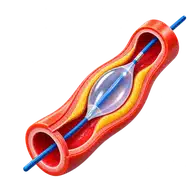
Under local anaesthesia, a catheter was guided from an artery in the groin to the uterine arteries. Embolic particles were delivered to reduce the fibroids’ blood supply. Pain relief and monitoring are important parts of care during and after UAE.
Reported recovery and follow-up
The original account records discharge the next day without further complications during that admission. At the one-week follow-up, the patient reported relief from pain and heavy bleeding. Longer-term symptom response and fibroid shrinkage require follow-up; a short-term improvement does not establish permanent relief.
Discussing suitability
Ask about post-embolisation pain, infection, changes in ovarian function, persistent symptoms and the chance of further treatment. Keeping the uterus does not guarantee preserved fertility. Anyone planning a pregnancy should discuss UAE and alternatives with the gynaecology and interventional teams.
For fibroid assessment in Vijayawada, bring ultrasound or MRI images, blood tests and previous treatment details. See the fibroid and UAE treatment guide or request an appointment.