Advanced minimally invasive care in Vijayawada

Author name: shankar

UTERINE ARTERY EMBOLISATION FOR FIBROIDS

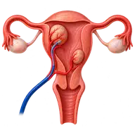
Uterine Artery Embolisation for Uterine Fibroids

Symptomatic uterine fibroids: the reported case A 35-year-old woman had pain and heavy bleeding for several months. The clinic’s original case account records uterine fibroids, with the largest measuring 6 cm. Following assessment, she chose uterine artery embolisation (UAE) as a treatment option. Imaging and planning The report describes ultrasound and MRI to assess fibroid […]

Uterine Artery Embolisation for Uterine Fibroids Read More »
PTBD and Metallic T- stenting

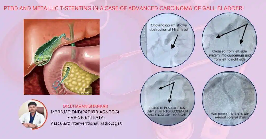
PTBD and Metallic T-Stenting in Advanced Carcinoma of Gall Bladder

Gall bladder carcinoma with obstructive jaundice The clinic’s report describes a 52-year-old man with advanced gall bladder carcinoma, obstructive jaundice, severe itching and loss of appetite. His initial total bilirubin was recorded as 16. The team planned percutaneous transhepatic biliary drainage (PTBD) and metallic T-stenting to improve bile drainage. Why drainage was considered A tumour

PTBD and Metallic T-Stenting in Advanced Carcinoma of Gall Bladder Read More »

A-V Fistuloplasty

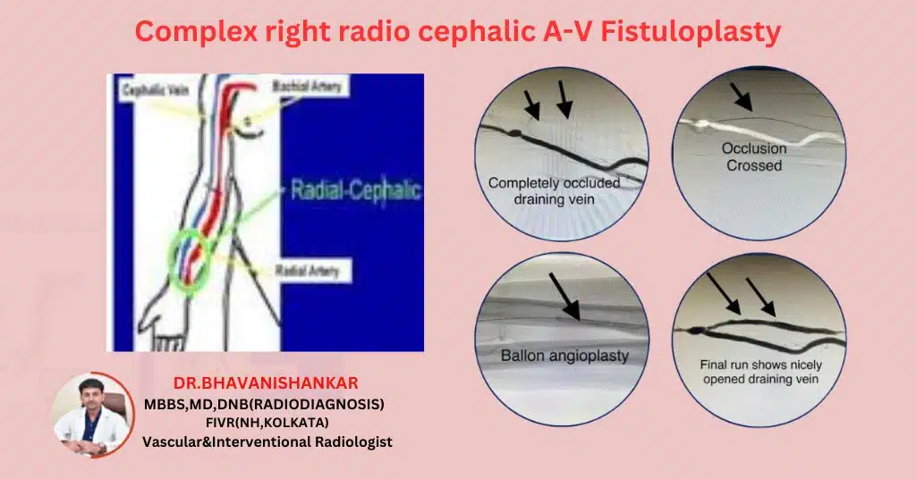
Complex right radio cephalic A-V Fistuloplasty

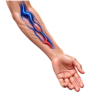
Right radiocephalic AV fistula narrowing The original clinic article describes a young patient receiving haemodialysis who developed narrowing of a right radiocephalic arteriovenous (AV) fistula. An AV fistula is the artery-to-vein connection used for dialysis access. Fistuloplasty is the procedure used to widen a narrowed section; it is not the name of the access itself.

Complex right radio cephalic A-V Fistuloplasty Read More »

fistula-chronic kidney disease

Fistula Wellness: Left Radio-Cephalic Fistuloplasty

Left radiocephalic dialysis fistula dysfunction The clinic’s case account describes a 45-year-old woman with chronic kidney disease whose left radiocephalic arteriovenous (AV) fistula was not providing adequate blood flow for dialysis. An AV fistula is the surgically created artery-to-vein connection used for haemodialysis access. Findings affecting the access Assessment identified a long segment of narrowing

Fistula Wellness: Left Radio-Cephalic Fistuloplasty Read More »

Uterine AVM

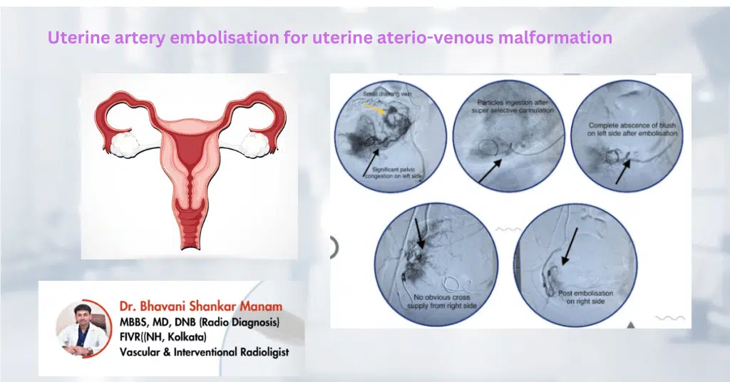
“UAE for Uterine AV Malformation”

Postpartum bleeding and suspected uterine vascular abnormality The original clinic report describes a 32-year-old woman with previous surgeries who developed recurrent bleeding after childbirth. Two months after delivery, she was experiencing intermittent episodes that were sometimes heavy. Imaging raised concern about an abnormal uterine blood vessel, and uterine artery embolisation was planned. Assessment before embolisation

“UAE for Uterine AV Malformation” Read More »

vic-Featured image

Mesenteric Aneurysm Embolization: A Lifesaver

Superior mesenteric artery aneurysm: presentation The clinic’s report describes a 57-year-old woman who presented with sudden abdominal pain. CT imaging identified an aneurysm of the superior mesenteric artery, a major vessel supplying the bowel. The symptoms and imaging prompted assessment for treatment. Why the artery’s anatomy matters An aneurysm is an abnormal enlargement of a

Mesenteric Aneurysm Embolization: A Lifesaver Read More »

PTBD AND METALLIC STENTING IN CASE OF CARCINOMA PANCREAS

PTBD and Metallic Stenting in Pancreatic Carcinoma Cases

Obstructive jaundice in pancreatic carcinoma Cancer in the head of the pancreas can obstruct the bile duct. Bile then cannot drain normally, causing jaundice, itching and sometimes poor appetite. Percutaneous transhepatic biliary drainage (PTBD) and metallic biliary stenting may help selected patients when drainage is needed. These procedures relieve obstruction; they do not remove or

PTBD and Metallic Stenting in Pancreatic Carcinoma Cases Read More »

Giant liver haemangiomas

Successful Management of Giant Liver Haemangioma through Transarterial Chemoembolization (TACE)

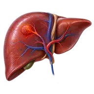
Giant liver haemangioma: the reported case A 65-year-old man presented with persistent abdominal pain and a feeling of fullness. Imaging identified a liver mass measuring 12 × 10 cm, described in the original clinic report as a giant liver haemangioma. A haemangioma is a benign vascular lesion and is different from liver cancer. Imaging and

Successful Management of Giant Liver Haemangioma through Transarterial Chemoembolization (TACE) Read More »

pulmonary artery pseudoaneurysm

Successful Pulmonary Artery Pseudoaneurysm Embolization

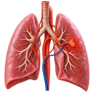
Pulmonary artery pseudoaneurysm with coughing blood The clinic’s case report describes a 73-year-old woman with tuberculosis who developed episodes of massive haemoptysis, or coughing up blood. Imaging identified a large pseudoaneurysm in a segmental pulmonary artery in the left lower lobe. This was assessed as the source of bleeding. Assessment and embolisation A pseudoaneurysm is

Successful Pulmonary Artery Pseudoaneurysm Embolization Read More »

Scroll to Top